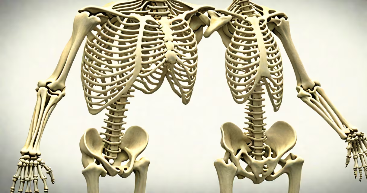
An appendicular skeleton diagram illustrates all bones in the limbs and their associated girdles-the pectoral (shoulder) and pelvic (hip) girdles-that attach the limbs to the axial skeleton. It shows the structural foundation of movement in the arms, legs, hands, and feet.
The appendicular skeleton includes 126 bones in adults-120 in the limbs themselves and six in the girdles. These bones enable walking, grasping, lifting, and fine motor control. The diagram separates the upper and lower limbs, highlighting the scapula, clavicle, humerus, radius, ulna, carpals, metacarpals, phalanges, pelvis, femur, tibia, fibula, tarsals, metatarsals, and phalanges.
Each limb begins at a girdle. The pectoral girdle connects the upper limbs to the sternum and thoracic vertebrae via the clavicles and scapulae. The pelvic girdle fuses the lower limbs to the sacrum through the hip bones (ilium, ischium, pubis). The diagram shows how these girdles serve as stable anchors for limb motion while allowing wide ranges of movement.
Muscles attach to bones in the appendicular skeleton via tendons. When muscles contract, they pull on bones, creating joint movement. For example, the biceps brachii contracts to flex the elbow, pulling the radius and ulna toward the humerus. A diagram labels these attachment points and lever systems.
Joint types determine movement. Ball-and-socket joints (like the shoulder and hip) allow rotation in multiple directions. Hinge joints (elbow and knee) permit flexion and extension. The diagram uses color coding or labeling to distinguish joint types and their degrees of freedom.
A single diagram may show both sides of the body. Left and right limbs mirror each other. This symmetry reflects evolutionary adaptation for bipedalism and bilateral dexterity. The left arm's humerus aligns with the right arm's in position and orientation, though mirrored.
Diagrams vary by purpose, detail level, and audience. Medical students see full-color, labeled diagrams with muscle attachments. Educators use simplified versions for middle school biology. Digital interactive models allow users to rotate, zoom, and isolate structures.
| Type | Detail Level | Use Case | Example |
|---|---|---|---|
| Basic | Minimal labels, outlines | Classroom introduction | Textbook figure showing limb segments |
| Standard | Full bone names, joint types | High school or college anatomy | Diagram in Gray's Anatomy reference |
| Interactive | 3D model, clickable parts | Medical training, virtual labs | Visible Body software or Netter's Atlas |
Some diagrams emphasize functional relationships. For instance, a diagram showing the kinetic chain during walking highlights how force travels. Others focus on pathology-such as dislocation patterns in the shoulder or fractures in the femoral shaft.
Many assume the appendicular skeleton includes the skull or spine. It does not. The axial skeleton-skull, vertebral column, ribs-supports the head and trunk. The appendicular skeleton only covers limbs and girdles.
Another error is thinking the pelvic girdle is a single bone. It is formed by three fused bones: ilium, ischium, and pubis. The diagram must show this fusion. A common mistake in older illustrations is drawing the hip bones as separate pieces.
A non-obvious fact: the clavicle is the only long bone in the body that lies horizontally. Most long bones run vertically. Its horizontal orientation allows the shoulder to rotate freely while maintaining stability. A well-labeled diagram shows this unique angle.
In a physical therapy clinic, a therapist uses an appendicular skeleton diagram to explain a patient's knee injury. The patient fell while skiing and tore the anterior cruciate ligament (ACL). The therapist points to the femur, tibia, and ACL on the diagram.
"The ACL stabilizes the knee," the therapist says. "When you twist suddenly, this ligament can snap. We'll use the diagram to show how the tibia moves forward under the femur when the ACL is damaged."
The therapist then compares the injured side to the healthy side using a split-screen diagram. The patient sees how the alignment shifts. This visual aid improves understanding and treatment compliance.
I once used a printed appendicular skeleton diagram during a high school science fair. Students had to identify bones in a puzzle format. One student struggled with the carpal bones. After pointing to the diagram's labeled wrist section, he correctly identified the scaphoid and lunate within 30 seconds. The visual cue made the difference.
Always verify diagram accuracy against peer-reviewed sources. According to Gray's Anatomy (41st edition), the standard terminology for limb bones follows the International Terminology of Anatomy (Terminologia Anatomica). Diagrams that use outdated terms like "wrist bone" instead of "carpal bone" are misleading.
When studying, cross-reference your appendicular skeleton diagram with clinical imaging. A CT scan of a fractured femur shows the same bones shown in the diagram. The correlation between visual representation and real anatomy builds lasting knowledge.
Every diagram is a tool. The most effective ones combine clarity, precision, and context. Whether used in classrooms, clinics, or research, a correct appendicular skeleton diagram enables accurate communication about human movement and structure.