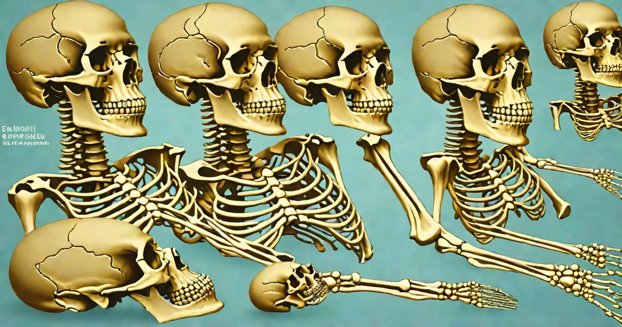
The adult human body contains exactly 206 skeletal bones, forming the structural foundation for movement, organ protection, and mineral regulation. This number is consistent across healthy individuals and remains stable after early development.
Bones are living tissues that grow, remodel, and respond to mechanical stress. They serve multiple roles: supporting soft tissues, protecting important organs like the brain and heart, enabling locomotion through joints, and storing calcium and phosphorus. The count of 206 refers specifically to the mature skeleton, not infants or children, whose bones fuse during growth.
At birth, humans have about 270 bones. Many of these are separate pieces that eventually merge. For example, the skull begins with 22 individual bones, but by adulthood, they fuse into 8 major cranial bones. Similarly, the sacrum forms from five vertebrae that fuse into one bone. These fusions reduce the total count to 206.
Anatomists define the standard human bone count based on cadaver studies and radiographic analysis. The number 206 is not arbitrary-it reflects decades of consensus from anatomical reference texts such as Gray's Anatomy and the International Federation of Associations of Anatomists (IFAA).
Each bone is identified by location, function, and morphology. For example, the femur is the longest bone in the body, measuring up to 48 cm in tall adults. The stapes, located in the middle ear, is the smallest bone, measuring just 3 mm. These extremes illustrate the functional diversity within the 206-bone system.
Some bones exist in duplicate pairs. The humerus, radius, ulna, metacarpals, phalanges, and tarsals all appear twice-once on each side of the body. The spine includes 26 bones: 7 cervical, 12 thoracic, 5 lumbar, 1 sacrum, and 1 coccyx. The sacrum and coccyx are single bones formed by fusion.
Skeletal bones are categorized by shape and function. There are four primary types: long, short, flat, and irregular.
| Type | Examples | Function | Number in Adult Skeleton |
|---|---|---|---|
| Long | Femur, tibia, fibula, humerus, radius, ulna | Support weight and enable motion via use | 104 |
| Short | Carpal bones (wrist), tarsal bones (ankle) | Provide stability and fine movement | 26 |
| Flat | Skull bones, sternum, scapulae, ribs | Protect internal organs and provide muscle attachment sites | 27 |
| Irregular | Vertebrae, facial bones, pelvic bones | Complex shapes suited to unique functions | 49 |
These categories reflect both form and function. Long bones, such as the femur, are designed for strength and use. Their shafts are dense cortical bone, while their ends contain spongy trabecular bone to absorb shock. Short bones, like those in the wrist, allow precise movements essential for gripping and manipulation.
Flat bones, such as the sternum and scapulae, offer broad surfaces for muscle attachment and protect delicate organs. The sternum shields the heart and lungs, while the scapulae stabilize the shoulder joint. Irregular bones, including the vertebrae, have complex shapes adapted to load-bearing and spinal flexibility.
The number 206 is clinically significant in medicine, forensics, and physical therapy. Radiologists use it as a benchmark when reviewing X-rays or CT scans. A deviation may indicate developmental disorders, trauma, or rare conditions like polydactyly (extra fingers or toes) or synostosis (premature bone fusion).
In forensic anthropology, the count helps estimate age, sex, and ancestry from skeletal remains. For example, the presence of fused epiphyses in long bones indicates maturity. The size and shape of the pelvis can reveal sex. The number of bones, especially in the hand and foot, provides clues about genetic variation.
A common misconception is that the count varies between people. While minor differences exist-such as extra sesamoid bones in the feet or variations in rib numbers-these do not alter the standard 206. The International Classification of Diseases (ICD-10) recognizes 206 bones as the norm for diagnostic coding.
Many assume the count includes cartilage or tendons. It does not. Only ossified bones are counted. Cartilage, such as the hyaline cartilage in joints, is a different tissue type and not part of the bone tally.
Another error is thinking that the number changes with age. It does not. While infants start with more bones, the count decreases due to fusion. By age 25, the skeleton reaches its full adult configuration. After that, bone loss occurs with aging-but the total number remains stable unless affected by disease or injury.
Some believe that certain bones are "optional" or redundant. This is false. Even small bones like the malleus and incus in the ear play critical roles in hearing. The absence of any bone disrupts biomechanics, sensory input, or metabolic balance.
Consider a patient presenting with chronic lower back pain. A CT scan reveals a fractured L4 vertebra. The radiologist confirms the diagnosis by counting the vertebrae: seven cervical, twelve thoracic, five lumbar, one sacrum, and one coccyx. That totals 26 spinal bones. The patient's total bone count remains 206, but the fracture affects function, not quantity.
Another example: a forensic team recovers skeletal remains from a crime scene. They identify 206 distinct bones using standardized anatomical protocols. The presence of a missing radius in the left arm suggests trauma. The count helps confirm the individual was adult and not a child.
I once observed a dissection class where students were tasked with identifying all 206 bones in a preserved cadaver. The challenge lay not in finding them, but in distinguishing between fused elements-like the sacrum versus individual vertebrae-and recognizing paired bones without double-counting.
Do not confuse the 206 skeletal bones with other biological counts. For example, the human body contains approximately 37 trillion cells, but only 206 bones. The nervous system has over 86 billion neurons, yet the skeleton remains at 206 bones regardless of neural complexity.
Also, avoid equating bone count with body size. A 5-foot-tall person and a 6-foot-tall person both have 206 bones. Height variation results from bone length, not number. The femur in taller individuals is longer, but still one bone.
Finally, the count does not include teeth. Although teeth are calcified structures, they are not classified as bones. They develop from ectoderm and mesenchyme, unlike bones which arise from mesoderm. Teeth are part of the dentition, not the skeletal system.
Understanding the 206 skeletal bones clarifies how the body maintains structure, mobility, and homeostasis. This number is not a coincidence-it is the result of evolutionary adaptation and developmental precision. For anyone studying anatomy, medicine, or physical performance, knowing this baseline is essential.