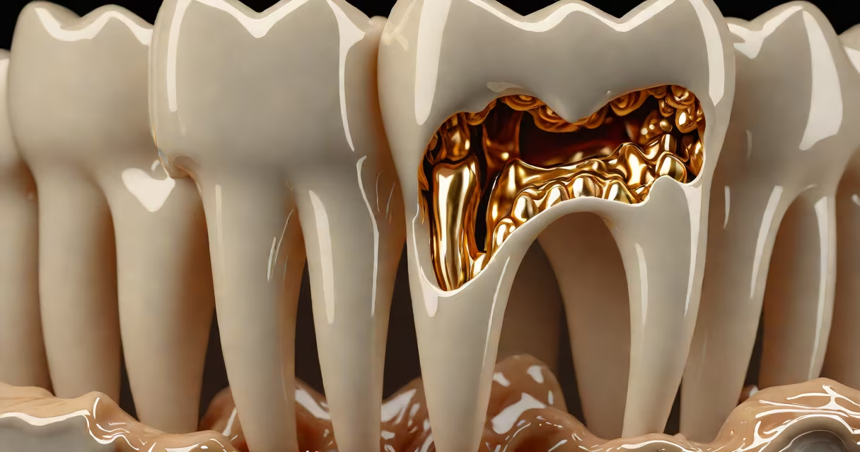
Cavities are small holes or structural breakdowns in teeth caused by bacterial acid erosion. They start as discolored spots and progress to visible pits or deep holes in enamel and dentin. Early-stage cavities may appear as brown or black stains, while advanced ones show visible openings or crumbling tooth structure.
Teeth develop cavities when plaque-a sticky film of bacteria-builds up on surfaces. These microbes feed on sugars from food and produce acid that dissolves tooth enamel. Over time, the acid wears through enamel and reaches the softer dentin layer beneath. Once dentin is affected, decay spreads faster and causes pain or sensitivity.
According to the American Dental Association (ADA), untreated cavities can lead to infections, abscesses, and tooth loss. The Centers for Disease Control and Prevention (CDC) reports that nearly 1 in 3 adults aged 20-64 have at least one untreated cavity. Early detection prevents irreversible damage and reduces treatment costs.
Cavities evolve through distinct visual stages. Recognizing them early improves outcomes. The earliest sign is a subtle change in tooth color-white, yellow, or brown spots on the surface. These areas feel slightly rough when touched with a tongue or dental probe.
As decay advances, the enamel begins to break down. A small pit or hole forms. This stage often appears as a darkened indentation, especially between teeth or along the gumline. The hole may trap food particles and feel sticky or gritty when probed.
In later stages, the cavity extends into dentin-the layer beneath enamel. The hole becomes larger, deeper, and may contain debris. Visible discoloration ranges from grayish-black to yellowish-brown. Pain during eating or drinking cold liquids indicates nerve involvement.
| Stage | Appearance | Common Location | Symptoms |
|---|---|---|---|
| Early | White or brown spots; rough texture | Top of molars, between teeth | None or mild sensitivity |
| Mild | Small pit or hole; dark staining | Gumline, chewing surfaces | Occasional pain with sweets |
| Advanced | Visible hole; crumbling tooth structure | Deep in grooves, near roots | Persistent pain, swelling |
Many people assume cavities always cause pain. That's false. Early cavities often show no symptoms. You might not notice a cavity until it grows large enough to affect nerves or cause visible damage.
Another myth is that only sugary foods cause cavities. While sugar fuels cavity-causing bacteria, acidic foods and drinks-like soda, citrus fruits, and energy drinks-also erode enamel. Acid weakens tooth structure, making it easier for bacteria to invade.
A lesser-known fact: some cavities form under existing fillings or crowns. These are called secondary or recurrent cavities. They're hard to see without dental X-rays because decay starts beneath the restoration material. The American Academy of Pediatric Dentistry notes that up to 15% of dental restorations fail within 10 years due to hidden decay.
I once noticed a dark spot on my lower molar while brushing. It was about the size of a pinhead and felt slightly rough. I didn't have pain, but I was concerned. I visited a dentist who used a dental explorer-a thin metal tool-to confirm a soft area in the tooth. An X-ray showed decay had reached the dentin layer. The dentist removed the damaged tissue and filled the hole.
The cavity wasn't visible to me without close inspection. I'd missed it for months because it lacked symptoms. This mirrors CDC findings: 40% of adults with cavities report no pain until the problem worsens. Regular dental checkups catch issues before they become serious.
Cavities are not the same as tooth cracks, enamel wear, or staining. Cracks appear as thin lines running through the tooth. Enamel wear shows as smooth, shiny patches. Stains result from food, tobacco, or medications-not bacterial decay.
Some people confuse white spots on teeth with early cavities. These can be signs of enamel demineralization-reversible with fluoride treatments. True cavities involve actual breakdown of tooth structure. A dentist can distinguish between them using tools like a dental probe or transillumination light.
Children often develop cavities on the front teeth due to frequent sipping of juice or soda. Early signs include chalky white patches on incisors. Parents may mistake these for normal tooth development. But they indicate early decay.
Adults face higher risks from dry mouth, which reduces saliva's protective effect. Saliva neutralizes acid and washes away food debris. Reduced flow increases cavity formation. People taking certain medications-like antihistamines or antidepressants-are more prone to decay.
When checking your mouth, look for changes in color, texture, or shape. Use a flashlight and mirror to examine back teeth. If you find a suspicious spot, don't wait. See a dentist. Early treatment saves time, money, and discomfort.
Understanding how cavities look like helps prevent complications. Regular brushing, flossing, and dental visits reduce risk. Fluoride strengthens enamel. Limiting sugary and acidic intake slows decay progression. Knowing what to watch for makes a lasting difference in long-term dental health.