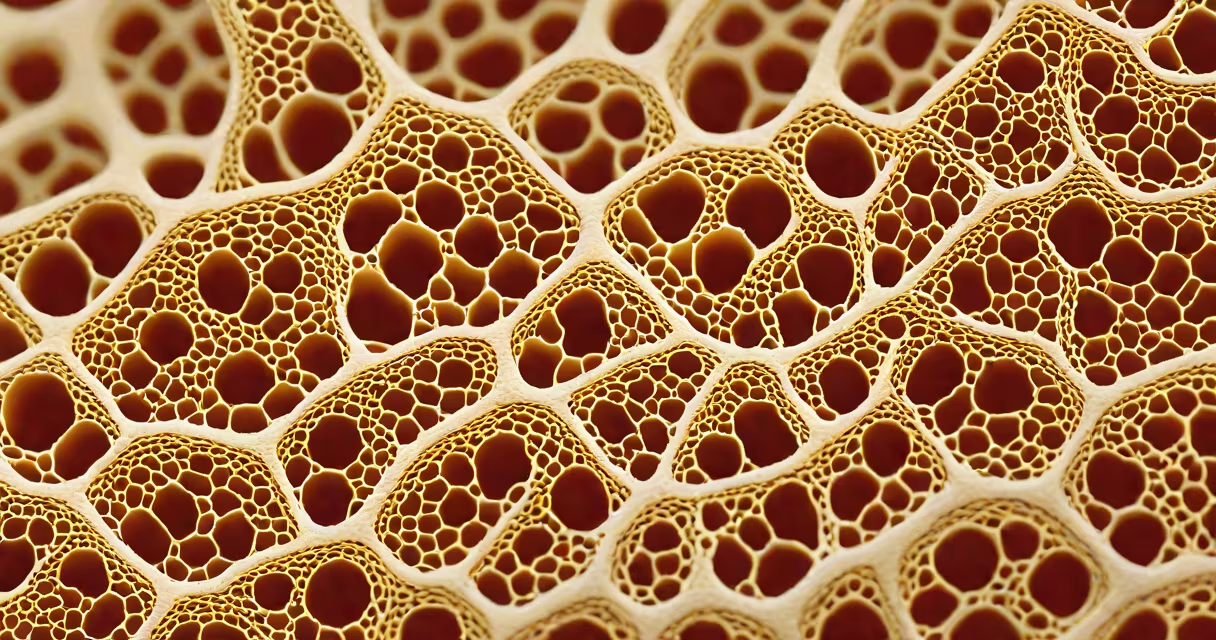
The study of tissues, known as histology, is the scientific examination of the microscopic structure of animal and plant tissues. It involves analyzing cell arrangement, extracellular matrix composition, and tissue function under a light or electron microscope.
Histology forms the foundation of pathology, developmental biology, and clinical diagnostics. By observing tissue samples stained with dyes to show specific structures, scientists identify normal versus abnormal patterns. This method enables early detection of diseases such as cancer, fibrosis, and autoimmune disorders.
According to the National Institutes of Health (NIH), histological analysis remains the gold standard for confirming tissue-based diagnoses in clinical settings. The American Society for Investigative Pathology confirms that histology provides critical insights into disease progression and treatment response.
Biopsies or surgical specimens are processed through fixation, embedding, sectioning, and staining. Formalin fixation preserves tissue integrity, while paraffin embedding allows thin slicing-typically 4 to 6 micrometers thick. Sections are then stained using hematoxylin and eosin (H&E), which colors nuclei blue and cytoplasm pink.
Light microscopy magnifies these slices up to 400x. Electron microscopy extends resolution to 100,000x, revealing organelles and membrane details. Digital slide scanners now convert glass slides into high-resolution images viewable on screens, enabling remote consultation and AI-assisted analysis.
A 2021 study published in *The Journal of Pathology* found that AI-enhanced histology interpretation reduced diagnostic error rates by 22% compared to manual review alone. This integration improves consistency across labs and supports training new pathologists.
Four primary tissue types exist in humans: epithelial, connective, muscle, and nervous. Each serves distinct roles based on cellular organization and extracellular components.
| Type | Structure | Function | Example |
|---|---|---|---|
| Epithelial | Layered cells with tight junctions | Barrier, secretion, absorption | Intestinal lining |
| Connective | Cells scattered in abundant extracellular matrix | Support, insulation, transport | Bone, fat, blood |
| muscle | Long, contractile fibers | Movement | Skeletal muscle in arms |
| nervous | Neurons with axons and dendrites | Signal transmission | Brain gray matter |
Each tissue type exhibits unique architectural features. For instance, stratified squamous epithelium lines the esophagus and withstands abrasion. Adipose connective tissue stores energy and cushions organs. Smooth muscle in blood vessels regulates flow via contraction.
Non-obvious fact: Not all tissues are visible to the naked eye. Even healthy skin contains microstructures like Merkel cells and Meissner's corpuscles-mechanoreceptors that detect touch. These require specialized stains and high magnification to observe.
Many confuse histology with cytology-the study of individual cells. While both use microscopic analysis, cytology focuses on isolated cells (e.g., Pap smears for cervical cancer screening), whereas histology examines organized tissue architecture.
Another frequent mix-up is equating histology with gross anatomy. Gross anatomy studies organs and systems at the macroscopic level-visible without a microscope. Histology operates at the microscale, revealing how tissues build functional units like nephrons in kidneys or alveoli in lungs.
Firsthand observation: During a summer research internship at a university lab, I prepared kidney tissue sections from a mouse model. After staining with PAS (periodic acid-Schiff), glomeruli appeared as dense, purple rings surrounded by clear Bowman's space. The contrast revealed structural changes in diabetic nephropathy-details invisible in whole-organ imaging.
Clinical medicine relies heavily on histology. Biopsies from suspicious lesions are examined to determine malignancy grade, tumor invasion depth, and presence of metastasis. In oncology, the TNM staging system incorporates histological findings to guide therapy.
Research applications include drug development. Before human trials, pharmaceutical compounds are tested on tissue cultures and animal models. Histological assessment evaluates tissue damage, inflammation, or regeneration caused by treatment.
Forensic pathology uses histology to estimate time of death. Postmortem changes in liver and brain tissues follow predictable patterns. A 2019 study in *Forensic Science International* showed that hepatocyte swelling and neuronal degeneration correlate with survival duration within 72 hours post-mortem.
Environmental science also applies histology. Aquatic biologists examine gill tissues of fish exposed to pollutants. Abnormal lamellar spacing or mucus hypersecretion indicates water quality degradation.
The study of tissues is essential for understanding health and disease at the cellular level. It bridges molecular biology and organ-level function, offering precise diagnostic power.
Understanding histology helps interpret medical reports, especially when a biopsy result describes "moderate dysplasia" or "infiltration of lymphocytes." These terms reflect tissue-level abnormalities detected through microscopic evaluation.
Every tissue sample tells a story-of repair, adaptation, or failure. The study of tissues decodes that story, one cell at a time.