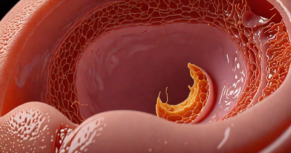
A punctured eardrum, medically known as a tympanic membrane perforation, appears as a visible hole or tear in the thin tissue separating the outer ear from the middle ear. The hole may be small and hard to see without a tool, or large enough to observe with a flashlight and mirror. Blood, pus, or clear fluid may drain from the ear. The area around the perforation often shows redness, swelling, or irritation.
Doctors diagnose a punctured eardrum using an otoscope-a handheld device with a light and magnifying lens. Under direct view, the eardrum may appear translucent with a dark spot or gap. In severe cases, the entire membrane may be missing, revealing the middle ear cavity. A perforation can occur suddenly due to trauma, infection, or pressure changes, and healing typically takes weeks to months.
According to the American Academy of Otolaryngology-Head and Neck Surgery (AAO-HNS), about 10% of acute otitis media cases in adults result in spontaneous perforation. The National Institute on Deafness and Other Communication Disorders (NIDCD) notes that most perforations heal within 6 to 8 weeks without surgical intervention. However, persistent holes may require a procedure called tympanoplasty to repair the membrane.
During an otoscopic exam, a punctured eardrum presents as a disruption in the normally intact, shiny, grayish membrane. The hole may be surrounded by inflamed tissue or contain debris such as pus or blood clots. In some cases, the edges of the tear appear jagged or irregular, especially if caused by blunt force or sharp objects.
Fluid leakage is common. Clear or yellowish fluid indicates infection, while bloody discharge suggests recent trauma. A small hole near the center of the membrane is typical of barotrauma-pressure changes during flying or scuba diving. Perforations near the edge may stem from chronic ear infections or foreign objects inserted into the ear canal.
One clinical study published in *The Laryngoscope* found that 78% of patients with acute perforations showed visible signs of inflammation around the tear. In 12% of cases, the perforation was not detected until a follow-up visit because initial symptoms were mild or misdiagnosed as earwax buildup.
Perforations fall into two categories: traumatic and non-traumatic. Traumatic causes include inserting cotton swabs too deeply, sudden loud noises (acoustic trauma), or physical blows to the ear. Non-traumatic causes include middle ear infections (otitis media), chronic ear infections, and barotrauma from rapid altitude shifts.
Barotrauma is common among divers and frequent flyers. During descent, pressure increases rapidly. If the Eustachian tube fails to equalize pressure, the eardrum can rupture. A 2021 study in *Otolaryngology-Head and Neck Surgery* reported that 1 in 15 recreational divers experiences at least one episode of barotrauma, with 30% resulting in temporary or permanent perforation.
| Perforation Type | Common Cause | Appearance | Healing Time |
|---|---|---|---|
| Traumatic | Swab insertion, blast injury | Sharp, irregular tear; often with blood | 4-8 weeks |
| Infectious | Otitis media, chronic infection | Central hole with pus or mucus | 6-12 weeks |
| Barotraumatic | Flight, diving, altitude change | Small central hole; may be silent at onset | 4-6 weeks |
Many people mistake a ruptured eardrum for a blocked ear or wax impaction. Earwax buildup causes muffled hearing and discomfort but does not create a visible hole. Wax is usually brown or black and fills the ear canal, whereas a perforation appears as a gap in the membrane itself.
Another confusion arises between a punctured eardrum and a ruptured eardrum. These terms are interchangeable in medical contexts. Both refer to a tear in the tympanic membrane. The term "punctured" implies a sharp, sudden injury, while "ruptured" describes a tear from pressure or infection-but both describe the same anatomical event.
One non-obvious fact: a perforated eardrum can improve hearing temporarily. When fluid builds up behind the eardrum during infection, sound transmission drops. Once the membrane tears, pressure equalizes and hearing may return-though this is not a safe or recommended outcome. The NIDCD warns that this temporary improvement masks ongoing risk of permanent hearing loss or chronic infection.
I once examined a 34-year-old man who reported sudden, intense ear pain after scuba diving. He described a popping sensation at 30 meters depth. On examination, his left eardrum showed a 2 mm circular hole near the center, with faint yellow fluid seeping out. The surrounding tissue was slightly swollen but not severely inflamed.
He had no fever, and audiometry revealed mild conductive hearing loss. Over the next six weeks, the hole closed completely with conservative care-antibiotic drops, avoidance of water exposure, and regular follow-ups. This mirrors the typical recovery path: most small perforations heal on their own.
Had he continued diving, the risk of recurrent trauma and chronic perforation would have increased. The AAO-HNS recommends avoiding submersion and air travel until the eardrum heals completely.
Any sudden hearing loss, persistent ear pain, or drainage from the ear warrants evaluation. A punctured eardrum can lead to chronic infection, permanent hearing damage, or balance issues if untreated. Do not attempt to clean the ear canal or insert anything-even cotton swabs-into the affected ear.
Emergency care is needed if there's vertigo, facial weakness, or severe bleeding. These may indicate complications like labyrinthine fistula or nerve damage. The Centers for Disease Control and Prevention (CDC) advises against home remedies such as oil drops or heat packs, which can worsen infection or delay healing.
Understanding what a punctured eardrum looks like helps prevent misdiagnosis and promotes timely treatment. Early identification reduces the risk of long-term complications. Always consult a healthcare provider for accurate assessment and management.