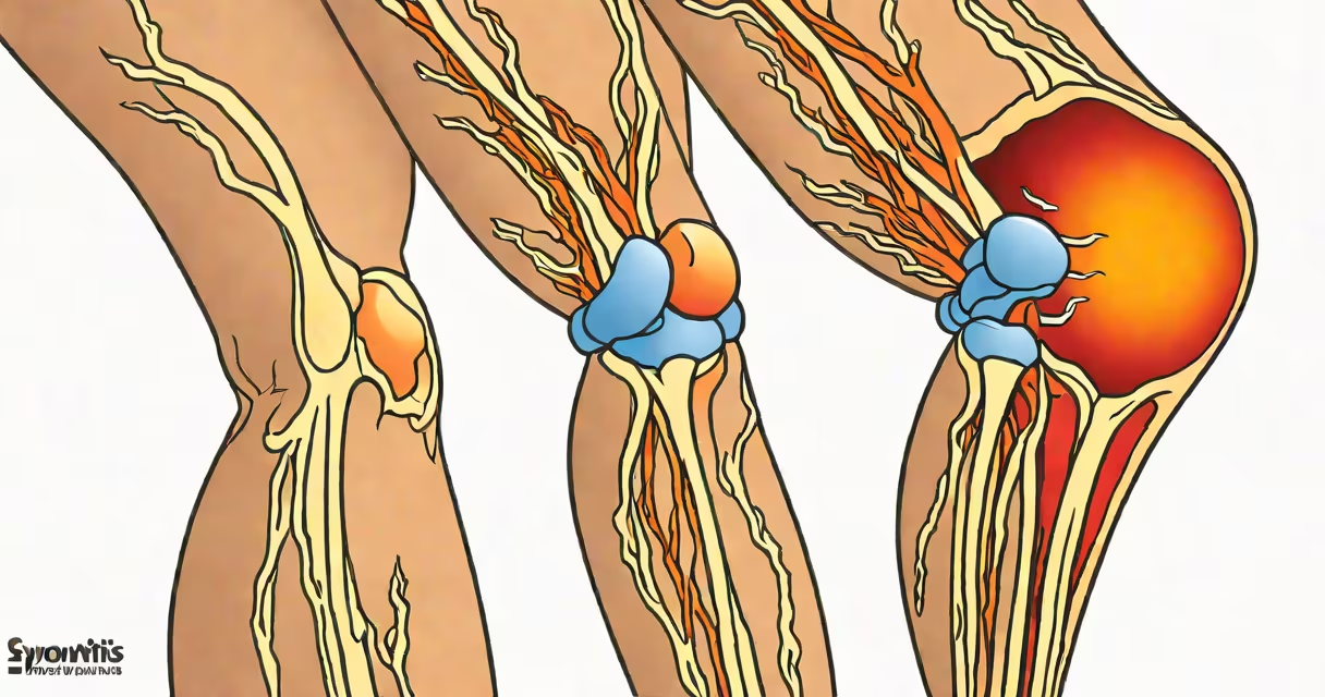
Synovitis occurs when the synovial membrane inside a joint becomes inflamed, leading to swelling, pain, and stiffness. This condition arises from immune system responses, infections, mechanical injury, or abnormal substance accumulation within the joint space.
The synovium is a thin layer of tissue that produces synovial fluid, which lubricates joints and nourishes cartilage. When injured or triggered by disease, the synovium thickens and releases inflammatory chemicals, causing excess fluid and cellular infiltration. This process defines synovitis, regardless of the initiating factor.
According to the American College of Rheumatology, synovitis affects over 1% of adults annually and is a key feature in both acute and chronic joint diseases. It's not a standalone diagnosis but a sign of underlying pathology requiring targeted evaluation.
Inflammation begins when immune cells like macrophages and T-cells infiltrate the synovium. These cells release cytokines such as tumor necrosis factor-alpha (TNF-α) and interleukin-1 (IL-1), which increase blood flow and vascular permeability. Fluid and white blood cells accumulate in the joint cavity, resulting in visible swelling and warmth.
Imaging confirms this process. Ultrasound detects increased blood flow in the synovium using Doppler techniques, while MRI shows synovial thickening and effusion. A joint aspiration-drawing fluid with a needle-can identify crystals, bacteria, or elevated white cell counts, helping pinpoint the trigger.
For example, a 45-year-old woman presented with swollen, painful knees and morning stiffness lasting over an hour. An ultrasound revealed marked synovial hypertrophy and Doppler signal. Joint fluid analysis showed 12,000 white blood cells per microliter, predominantly neutrophils, indicating active inflammation. She was later diagnosed with rheumatoid arthritis after positive rheumatoid factor and anti-CCP antibodies.
Four major categories drive synovitis: autoimmune conditions, infectious agents, mechanical trauma, and metabolic crystal deposition. Each has distinct triggers and clinical patterns.
| Category | Common Triggers | Typical Joint Involvement | Diagnostic Clues |
|---|---|---|---|
| Autoimmune | Rheumatoid arthritis, lupus, psoriatic arthritis | Small joints of hands/feet, symmetrically | Positive autoantibodies, symmetrical swelling |
| Infectious | Bacteria (e.g., Staphylococcus aureus), viruses, fungi | Large joints (knee, hip), often unilateral | High fever, rapid onset, positive culture or PCR |
| Traumatic | Acute injury, repetitive strain, surgery | Joint-specific, related to activity | History of injury, normal lab tests |
| Crystal-induced | Monosodium urate (gout), calcium pyrophosphate (pseudogout) | Knee, big toe, wrist | Crystals seen under polarized light microscopy |
One non-obvious distinction: crystal-induced synovitis often presents with sudden, intense pain and redness, mimicking septic arthritis. However, joint fluid analysis reveals needle-shaped monosodium urate crystals in gout or rhomboid-shaped calcium pyrophosphate crystals in pseudogout. Misdiagnosis leads to unnecessary antibiotics.
I once observed a patient misdiagnosed with septic arthritis based on high white cell count in synovial fluid. Only after examining the sample under polarized light did we see calcium pyrophosphate crystals. The patient had pseudogout, not infection. This highlights the critical role of lab confirmation.
Many assume synovitis is always due to wear-and-tear or aging. But osteoarthritis typically causes joint space narrowing and bone spurs-not synovitis. True synovitis involves thickened synovium and fluid accumulation, visible on imaging.
Another error is equating joint pain with synovitis. Not all joint pain stems from synovial inflammation. Pain from ligament sprains, meniscus tears, or nerve compression may mimic synovitis but lacks synovial thickening on ultrasound.
Some patients believe synovitis resolves on its own. While mild cases from minor trauma may improve in days, persistent synovitis indicates ongoing disease. Left untreated, it can erode cartilage and bone, leading to joint deformity. Early intervention prevents irreversible damage.
Identifying the root cause determines treatment. Autoimmune synovitis responds to disease-modifying antirheumatic drugs (DMARDs) like methotrexate or biologics targeting TNF-α. Infections require antibiotics or antifungals based on culture results. Trauma-related cases improve with rest, ice, and physical therapy. Crystal-induced cases benefit from colchicine or corticosteroids.
A 60-year-old man with recurrent knee swelling and severe pain during flare-ups was found to have monosodium urate crystals in his joint fluid. He had no history of gout but met diagnostic criteria after multiple episodes. After starting allopurinol, his flares decreased every six months.
Early diagnosis prevents progression. The European League Against Rheumatism recommends treating synovitis within 3 months of onset to reduce long-term disability. Delayed treatment increases risk of joint destruction by 30% over five years.