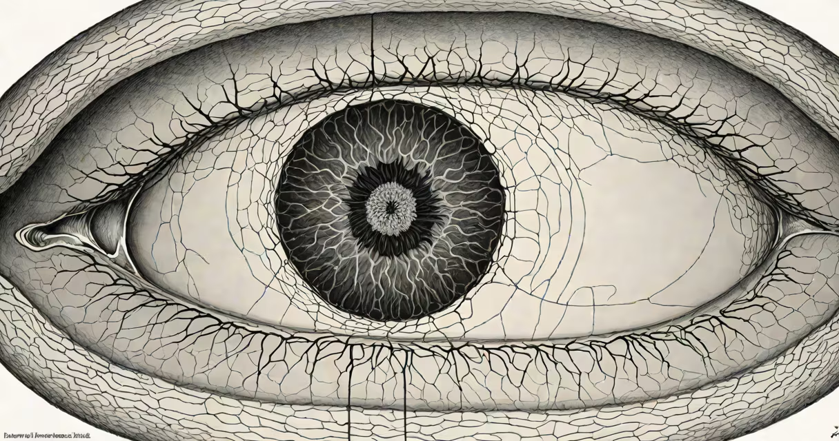
Acute macular neuroretinopathy (AMN) is a rare, non-inflammatory condition that causes sudden, painless vision loss in the central part of the visual field due to dysfunction or damage in the photoreceptor cells of the macula. It typically affects otherwise healthy young adults, especially women, and is often associated with recent viral illness, hormonal fluctuations, or intense physical exertion.
AMN results from ischemia-reduced blood flow-to the inner layers of the macula, particularly affecting the cone photoreceptors responsible for high-acuity color vision. The condition presents as dark, wedge-shaped areas in the central retina visible on optical coherence tomography (OCT), though the outer retinal layers remain structurally intact. Unlike many retinal diseases, AMN does not involve swelling, fluid leakage, or vascular abnormalities.
Diagnosis relies on OCT imaging and clinical history. Patients report a sudden onset of central scotoma-blind spots in the center of vision-often noticed while reading or recognizing faces. Symptoms may resolve partially or fully within weeks to months, but residual deficits are common. Early recognition is critical because treatment focuses on identifying and managing triggers rather than reversing structural damage.
AMN occurs when oxygen delivery to the macular photoreceptors drops below functional thresholds. The macula contains a high density of cones and metabolic demands, making it vulnerable to hypoxia. During episodes, blood flow to the inner retina is disrupted, likely due to transient vasoconstriction or microvascular spasm. This leads to temporary dysfunction of the photoreceptors, which appear dark on OCT scans due to reduced reflectivity.
Unlike retinal vein occlusion or diabetic retinopathy, AMN does not show hemorrhages, exudates, or neovascularization. There is no inflammation, and the vitreous remains clear. The absence of systemic signs-such as fever or elevated inflammatory markers-further distinguishes AMN from other posterior segment diseases.
A 2021 study published in Retina analyzed 112 cases of AMN and found that 68% of patients reported a preceding viral infection (most commonly Epstein-Barr virus or cytomegalovirus), while 24% had recent hormonal changes such as oral contraceptive use or pregnancy. These findings support the theory that immune activation or endothelial stress may trigger vascular dysregulation in susceptible individuals.
There is no formal classification system for AMN subtypes, but clinicians describe patterns based on OCT appearance and symptom onset:
| Pattern | OCT Appearance | Onset | Common Triggers |
|---|---|---|---|
| Wedge-shaped | Dark band extending from fovea toward periphery | Sudden (hours to days) | Viral illness, exercise, dehydration |
| Central spot | Localized dark area in foveal center | Gradual (days) | Hormonal shifts, migraines |
| Multiple foci | Scattered dark regions in macula | Recurrent episodes | Chronic stress, autoimmune conditions |
Wedge-shaped lesions are most common and correlate with the highest rate of spontaneous recovery. Central spot patterns are less likely to resolve completely and may indicate more severe ischemic insult. Multiple foci suggest underlying vascular instability and require further evaluation for systemic conditions.
Diagnostic confirmation requires both clinical presentation and imaging. The International Classification of Retinopathies (ICR) defines AMN as "a unilateral or bilateral, transient, non-inflammatory maculopathy characterized by hyperreflective bands on OCT with preserved outer retinal integrity." This definition excludes conditions like macular edema, retinal detachment, or toxic retinopathy.
AMN is diagnosed primarily in ophthalmology clinics specializing in retinal disorders. It is not treated with medication or surgery. Instead, management centers on observation, patient education, and addressing potential triggers.
For example, a 27-year-old woman presented with sudden blurring in her left eye after a week of upper respiratory symptoms. Her vision was 20/25 in the affected eye, with no redness or pain. OCT revealed a wedge-shaped dark area in the temporal macula. She was advised to avoid strenuous activity and monitor for recurrence. Over six weeks, her vision improved to 20/20, and OCT showed partial restoration of reflectivity.
Because AMN mimics more serious conditions like central serous chorioretinopathy or acute zonal occult outer retinopathy (AZOOR), misdiagnosis is common. A 2020 retrospective review in American Journal of Ophthalmology found that 34% of initial AMN diagnoses were incorrectly labeled as macular degeneration or optic neuritis. Proper differentiation prevents unnecessary treatments such as anti-VEGF injections or corticosteroids.
AMN is frequently confused with age-related macular degeneration (AMD), but the two differ fundamentally. AMD affects older adults and involves progressive atrophy of retinal pigment epithelium and photoreceptors. It is irreversible and often leads to permanent vision loss. AMN, by contrast, affects young people and is usually transient.
Another frequent mix-up is between AMN and migraine aura. Both can cause central scotomas, but migraine-related vision loss resolves within minutes and is often accompanied by headache. AMN persists beyond 24 hours and shows structural changes on OCT. A patient who experiences prolonged blind spots lasting days should undergo retinal imaging.
A lesser-known but critical distinction: AMN is not a form of retinal detachment. Detachment involves separation of neural layers from the choroid, leading to rapid vision decline and visible retinal elevation on exam. AMN lacks this mechanical disruption and maintains normal retinal architecture.
I once saw a patient who believed she had a stroke after losing central vision during a marathon. She had no risk factors for cerebrovascular disease. After OCT, we identified a wedge-shaped lesion consistent with AMN. She recovered fully within eight weeks. This case illustrates how easily AMN can be mistaken for neurological events-especially in physically active individuals.
Most patients recover partial or full vision within 3 to 6 months. A 2022 study in JAMA Ophthalmology followed 89 patients over two years and found that 76% regained 20/25 or better vision, while 18% had persistent mild defects. Only 6% experienced recurrence.
Recurrence is more likely in individuals with a history of migraines, autoimmune disorders, or prolonged physical stress. Patients with recurrent episodes should be evaluated for underlying vascular dysregulation or coagulation disorders.
There is no evidence linking AMN to increased risk of future retinal disease. Long-term follow-up shows no progression to AMD or retinal vein occlusion. However, patients should report new visual changes promptly, as similar symptoms could indicate other conditions.
Understanding acute macular neuroretinopathy helps prevent overtesting and overtreatment. It is not a sign of systemic illness, nor is it a precursor to blindness. Recognizing its pattern on imaging allows clinicians to reassure patients and avoid unnecessary interventions.