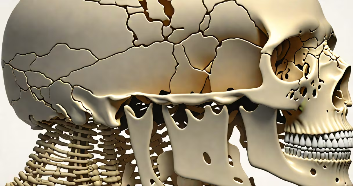
The anatomy of the axial skeleton refers to the central core of the human skeleton, including the skull, vertebral column, and thoracic cage. These bones form the body's axis and protect critical organs such as the brain, spinal cord, and heart.
This structure accounts for 80 bones in the adult human body-about half of the total skeletal count. The axial skeleton supports the head, maintains upright posture, and provides attachment points for muscles involved in breathing and neck movement.
Unlike the appendicular skeleton, which includes limbs and girdles, the axial skeleton is centralized and symmetrical. Its design prioritizes protection and stability over mobility. For example, the skull encases the brain, while the vertebral column houses the spinal cord and allows controlled bending and twisting.
Bones in the axial skeleton work together through joints, ligaments, and muscle attachments. The skull connects to the spine via the atlas (C1 vertebra), allowing nodding motion. The cervical, thoracic, and lumbar regions of the spine provide structural support and flexibility.
The thoracic cage-formed by the sternum, ribs, and thoracic vertebrae-protects the heart and lungs. During inhalation, the diaphragm contracts, increasing chest volume. The ribs move upward and outward, expanding the thoracic cavity. This motion is coordinated by intercostal muscles attached to the rib surfaces.
A real-world example: during a physical exam, doctors assess the alignment of the vertebral column using palpation and imaging. A misalignment in the lumbar region may cause sciatica due to nerve root compression. This highlights how precise bone positioning affects neurological function.
The axial skeleton consists of four main parts: cranial bones, facial bones, vertebral column, and thoracic cage. Each group serves distinct roles in protection, support, and movement.
| Component | Number of Bones | Key Functions |
|---|---|---|
| Cranial bones | 8 | Protect the brain; house sensory organs |
| Facial bones | 14 | Form face structure; support nasal passages and eyes |
| Vertebral column | 26 (including sacrum and coccyx) | Supports head; protects spinal cord; enables posture control |
| Thoracic cage | 25 (sternum + 24 ribs + 11 pairs of costal cartilages) | Protects heart and lungs; aids respiration |
Among these, the vertebral column is divided into five regions: cervical (7 vertebrae), thoracic (12), lumbar (5), sacral (5 fused), and coccygeal (4 fused). The cervical vertebrae allow head rotation, while the lumbar vertebrae bear most of the body's weight.
A common misconception is that the sacrum is a single bone. It is actually formed by five fused vertebrae. This fusion occurs between ages 16 and 25, contributing to pelvic stability during walking and childbirth.
Many assume the ribcage is rigid. In reality, it is flexible. Ribs articulate with the sternum via costal cartilage, which absorbs shock during running or jumping. Without this flexibility, breathing would be painful and inefficient.
Another error is equating the skull with the cranium. The cranium refers only to the eight bones enclosing the brain-the frontal, parietal (two), temporal (two), occipital, sphenoid, and ethmoid bones. The facial bones (like the maxilla and mandible) are separate and do not contribute to brain protection.
During my first dissection lab, I was surprised to see how much variation exists in the size and shape of the atlas vertebra. One student's specimen had a larger foramen magnum than average, affecting the head's balance point. This demonstrated that even small anatomical differences impact biomechanics.
In clinical practice, understanding the anatomy of the axial skeleton is essential for diagnosing conditions like scoliosis, kyphosis, and herniated discs. Scoliosis involves lateral curvature of the spine exceeding 10 degrees. It often develops during adolescence and may require bracing or surgery if progression continues.
According to the National Institute of Arthritis and Musculoskeletal and Skin Diseases (NIAMS), approximately 2 million people in the U.S. are diagnosed with osteoporosis annually. This condition weakens trabecular bone in the vertebrae, increasing fracture risk-even without trauma. Spinal fractures reduce height and alter posture.
For daily life, proper posture reduces strain on the axial skeleton. Sitting with a straight spine minimizes pressure on the lumbar discs. Research from the American Physical Therapy Association shows that poor sitting habits increase the risk of chronic back pain by 30% over ten years.
The anatomy of the axial skeleton is foundational to human biology. It governs protection, movement, and structural integrity. Mastery of its components enables accurate diagnosis, effective treatment, and deeper understanding of human form and function.