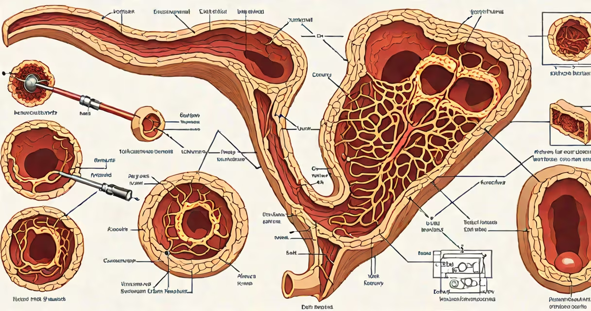
Erythematous mucosa gastric antrum refers to red, inflamed tissue observed in the antral region of the stomach during upper endoscopy. This appearance signals underlying mucosal injury, most commonly due to chronic inflammation from Helicobacter pylori infection, NSAID use, or autoimmune processes. The finding is not diagnostic on its own but prompts targeted biopsy and histopathological evaluation.
The gastric antrum is the lower portion of the stomach, near the pylorus. Its mucosa normally appears pale pink with visible vascular patterns. When inflamed, the surface becomes hyperemic-reddened due to increased blood flow-and may show loss of normal rugal architecture. Endoscopists describe this as erythematous, sometimes with patchy or diffuse involvement. The term "erythematous" specifically denotes redness caused by dilated capillaries and increased vascularity in the superficial mucosa.
A 2021 study published in Gastroenterology found that 68% of patients with endoscopic erythema in the antrum had histologically confirmed chronic gastritis upon biopsy. This confirms that visual assessment alone cannot distinguish between reactive changes and true inflammatory disease. Without tissue sampling, clinicians risk missing H. pylori colonization, which occurs in 30-50% of cases with antral erythema.
During esophagogastroduodenoscopy (EGD), the endoscopist evaluates the mucosa using high-resolution imaging. Erythema in the antrum is detected visually, often alongside other signs such as edema, friability, or erosions. The presence of erythema triggers targeted biopsies-typically four samples from the antrum and two from the corpus-to assess cellular infiltration and rule out malignancy.
Biopsy specimens are stained with hematoxylin and eosin (H&E) and assessed under a microscope. Pathologists look for lymphoid aggregates, plasma cell infiltrates, and neutrophil activity. A diagnosis of chronic gastritis requires sustained lymphoplasmacytic infiltration in the lamina propria. The updated Sydney classification system grades severity based on the extent of inflammation and atrophy.
For example, a patient presenting with epigastric discomfort undergoes EGD. The endoscopist notes diffuse erythema in the antrum, with loss of normal rugal folds. Four antral biopsies are taken. Histology reveals dense lymphoplasmacytic infiltrate, mild glandular atrophy, and H. pylori organisms identified via Giemsa stain. The final report states: "Chronic active gastritis, H. pylori-associated, moderate." This confirms the clinical significance of the endoscopic finding.
Not all erythematous mucosa in the gastric antrum stems from the same cause. The most common etiologies fall into three categories: infectious, drug-induced, and autoimmune.
| Category | Primary Cause | Prevalence in Antral Erythema | Key Clinical Clues |
|---|---|---|---|
| Infectious | Helicobacter pylori | 40-60% | Patchy erythema, nodularity, positive urease test |
| Drug-induced | NSAIDs, aspirin, bisphosphonates | 20-30% | Diffuse erythema, erosions, history of medication use |
| Autoimmune | Anti-parietal cell antibodies | 5-10% | Corpus-predominant atrophy, pernicious anemia risk |
One non-obvious distinction is that H. pylori-associated gastritis typically shows greater inflammation in the antrum than the corpus-a pattern known as "antral-predominant" gastritis. This contrasts with autoimmune gastritis, which primarily affects the corpus and leads to achlorhydria. Misidentifying the pattern can delay appropriate treatment.
I once observed a 52-year-old man with persistent dyspepsia. Endoscopy showed marked erythema in the antrum, but no ulcers. Biopsies revealed lymphoid follicles and neutrophils-consistent with chronic active gastritis. Testing was positive for H. pylori antigen in stool. After triple therapy (clarithromycin, amoxicillin, omeprazole), follow-up endoscopy six weeks later showed resolution of erythema. This case illustrates that erythematous mucosa is a marker, not a diagnosis.
Erythematous mucosa gastric antrum is frequently mistaken for acute ulceration or early neoplasia. However, the key difference lies in morphology. Ulcerations appear as crater-like defects with irregular borders. Neoplastic lesions often have nodular, polypoid, or flat elevations. Erythema presents as diffuse redness without structural disruption.
Another frequent error is assuming that erythema equates to H. pylori infection. While H. pylori is the leading cause, up to 35% of cases occur in H. pylori-negative individuals. Factors like bile reflux, smoking, and stress contribute to mucosal irritation. A 2020 review in Alimentary Pharmacology & Therapeutics noted that 28% of patients with antral erythema had no identifiable pathogen after complete testing.
Some endoscopists rely too heavily on visual cues. A 2019 multicenter study found that interobserver agreement for diagnosing erythema was only 62%. Variability increases when erythema is subtle or masked by mucus. This highlights the necessity of biopsy-especially in patients with persistent symptoms despite negative serology.
When erythematous mucosa gastric antrum is identified, the standard pathway includes histological analysis, H. pylori testing, and symptom assessment. If H. pylori is confirmed, eradication therapy is initiated. Guidelines from the American Gastroenterological Association recommend testing for H. pylori in all patients with chronic gastritis, regardless of symptom severity.
If biopsy shows mild chronic inflammation without active infection, the condition may be managed conservatively. Patients are advised to avoid NSAIDs, reduce alcohol intake, and monitor for worsening symptoms. Follow-up endoscopy is recommended if dyspepsia persists beyond eight weeks.
Erythematous mucosa gastric antrum is a signpost, not a destination. It signals the need for deeper investigation. Without biopsy and correlation with clinical context, it remains a descriptive observation with limited predictive power.