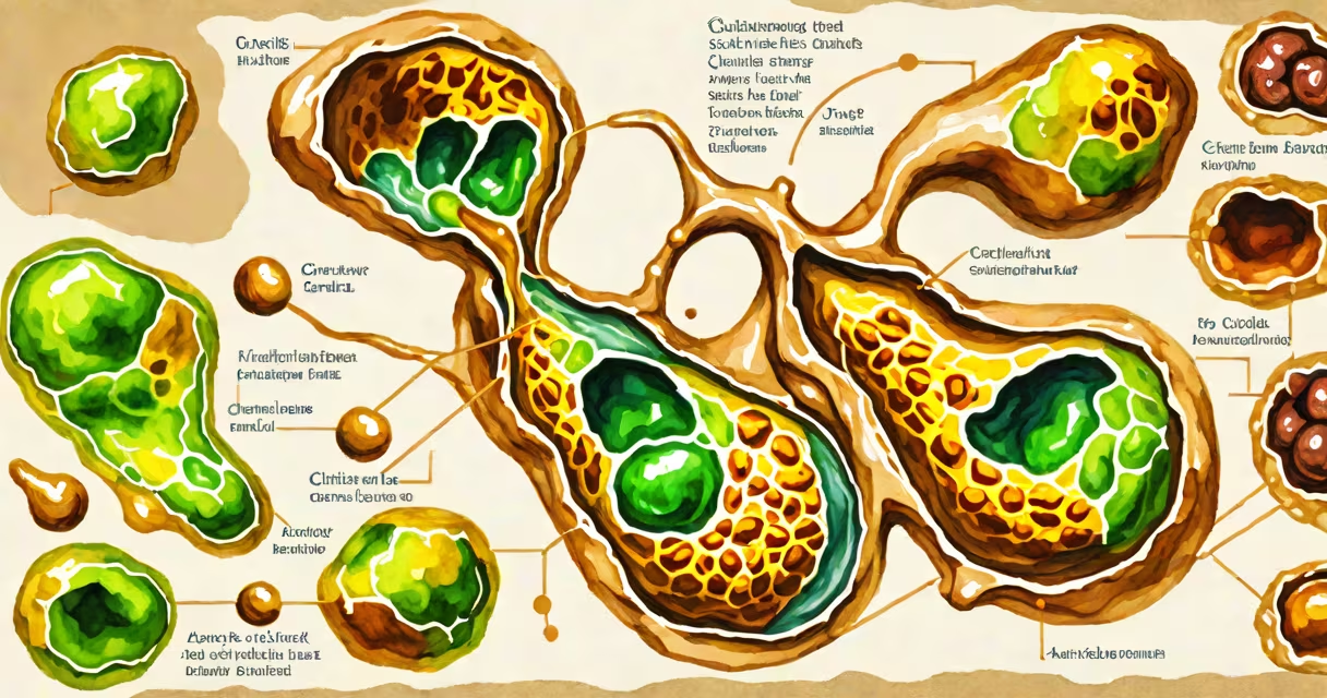
Gallstones form when substances in bile-primarily cholesterol or bilirubin-crystallize into solid particles inside the gallbladder. These stones can remain asymptomatic or cause severe pain when blocking ducts. The condition affects about 10-15% of adults in the U.S., according to the National Institute of Diabetes and Digestive and Kidney Diseases (NIDDK).
Bile, produced by the liver and stored in the gallbladder, aids fat digestion. When bile composition becomes imbalanced-either too much cholesterol or insufficient bile salts-cholesterol crystals can form. Pigment stones, less common, develop from excess bilirubin, often in people with liver disease or hemolytic anemia.
Most gallstones are small-about 6 mm in diameter-but can grow larger. They may stay in the gallbladder for years without causing issues. Once they move into the cystic or common bile duct, they trigger acute biliary colic, a sharp pain in the upper right abdomen lasting 1-5 hours.
Cholesterol gallstones form when the liver secretes bile with too much cholesterol relative to bile salts and lecithin. The imbalance causes cholesterol to precipitate out of solution. Over time, these microscopic crystals aggregate into stones. This process typically takes months to years.
Pigment gallstones result from elevated unconjugated bilirubin levels. Bilirubin forms when red blood cells break down. Conditions that increase red blood cell turnover-such as sickle cell disease or hereditary spherocytosis-raise bilirubin output. When the liver cannot process it efficiently, bilirubin precipitates and forms dark, granular stones.
A key factor is gallbladder motility. If the gallbladder doesn't empty properly, bile stagnates. Stagnant bile increases the chance of crystal formation. This is why prolonged fasting or rapid weight loss can trigger stone development-both reduce gallbladder contractions.
People with certain traits are more likely to develop gallstones. The NIDDK identifies four major risk factors: female sex, age over 40, obesity, and family history. Women are twice as likely as men to develop symptomatic gallstones, partly due to estrogen's effect on increasing cholesterol secretion into bile.
Obesity increases risk by 20-30%. Excess body fat alters bile composition and reduces gallbladder emptying. Rapid weight loss-especially losing more than 1.5 kg (3.3 lbs) per week-can double the likelihood of stone formation. A 2018 study in Journal of Hepatology found that participants who lost weight quickly had a 2.3-fold higher incidence of gallstones compared to gradual losers.
Other triggers include pregnancy, high-dose estrogen therapy (like oral contraceptives), and diets high in saturated fats and refined carbohydrates. A diet low in fiber also contributes. For example, a 2020 analysis of the NHANES dataset linked low dietary fiber intake to a 40% increased risk of gallstone disease.
| Type | Composition | Prevalence | Common Causes |
|---|---|---|---|
| Cholesterol | 90% of cases; mostly cholesterol monohydrate | ~75% | High cholesterol in bile, poor gallbladder emptying |
| Pigment (black) | Calcium bilirubinate; associated with chronic hemolysis | ~15% | Liver disease, hemolytic disorders |
| Pigment (brown) | Calcium salts of fatty acids; linked to infection | ~10% | Biliary tract infections, stasis |
Many assume gallstones are caused by eating greasy food. While fatty meals can trigger pain in people with existing stones, they don't cause stone formation. The misconception persists because the pain-often felt after a rich meal-is mistaken for the root cause.
Another error is equating gallstones with gas. People frequently confuse biliary colic with intestinal gas pain. Gas-related discomfort tends to be lower in the abdomen and may involve bloating or flatulence. Gallstone pain is localized to the upper right quadrant, often radiates to the right shoulder, and lasts longer than typical gas cramps.
Gallstones are not contagious or infectious. They do not spread between people. The condition arises from internal metabolic processes, not pathogens. This distinguishes them from conditions like hepatitis or cholangitis, which involve infection and inflammation of the bile ducts.
A 48-year-old woman with a BMI of 34 presented with sudden onset of severe right upper quadrant pain lasting 3 hours. She reported no fever, jaundice, or nausea. Her medical history included type 2 diabetes and a 20-pound weight loss over six weeks using a very-low-calorie diet.
An ultrasound revealed multiple gallstones, including a 1.2 cm stone lodged in the cystic duct. Blood tests showed normal liver enzymes and bilirubin. The diagnosis was symptomatic cholesterol gallstones. She underwent laparoscopic cholecystectomy and recovered within seven days.
This case illustrates how rapid weight loss and metabolic changes combine to increase risk. The patient had no prior symptoms, highlighting that gallstones can remain silent until they obstruct flow.
Not all gallstones require treatment. Asymptomatic stones-found incidentally during imaging for other reasons-are usually monitored. But if a person experiences recurrent episodes of biliary colic, surgery is recommended.
Diagnosis begins with ultrasound, the gold standard. It detects stones with 95% accuracy. If ultrasound is inconclusive, magnetic resonance cholangiopancreatography (MRCP) provides detailed images of the biliary tree. Blood tests assess liver function and rule out infection or obstruction.
I've seen patients misdiagnose gallstones as heartburn or acid reflux. One individual took proton-pump inhibitors for months before an abdominal ultrasound confirmed gallstones. The pain resolved after surgery. This underscores the importance of accurate imaging when symptoms persist.
Understanding how people get gallstones clarifies a common but misunderstood condition. The process involves bile chemistry, gallbladder function, and lifestyle factors. Recognizing symptoms early improves outcomes and prevents complications like cholecystitis or pancreatitis.