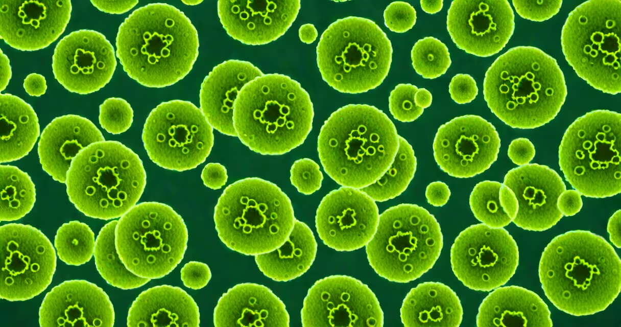
Streptococcus pneumoniae hemolysis refers to the partial destruction of red blood cells in blood agar cultures, resulting in a greenish or brownish discoloration around colonies. This pattern, known as alpha-hemolysis, is a defining characteristic used in laboratory identification of the bacterium.
Alpha-hemolysis occurs when hydrogen peroxide and other oxidizing agents produced by the bacteria partially degrade hemoglobin in red blood cells. The breakdown product, methemoglobin, gives the surrounding medium a greenish tint. This differs from beta-hemolysis, which results in complete lysis and clear zones, and gamma-hemolysis, which shows no change.
Streptococcus pneumoniae is a Gram-positive coccus that typically appears in pairs (diplococci) under the microscope. Its hemolytic pattern on blood agar-specifically alpha-hemolysis-is a critical early clue in distinguishing it from other streptococcal species such as group A streptococci (beta-hemolytic) or viridans group streptococci (also alpha-hemolytic but less consistently so).
The mechanism behind alpha-hemolysis in Streptococcus pneumoniae involves the production of hydrogen peroxide during metabolism. This reactive oxygen species oxidizes hemoglobin to methemoglobin, which is not fully degraded. The result is a visible greenish halo around the colony, often described as "greenish" or "brownish" depending on the culture's age and conditions.
This process is not destructive to the cell membrane. Red blood cells remain intact but change color due to altered iron states in the heme group. The reaction is temperature-sensitive and may vary slightly between strains, but consistent alpha-hemolysis remains a sign of S. pneumoniae on blood agar.
For accurate diagnosis, laboratories use multiple tests. Alpha-hemolysis alone is not sufficient to confirm S. pneumoniae. Other methods, including optochin sensitivity testing and bile solubility, are required to differentiate it from similar organisms.
Different streptococcal species exhibit distinct hemolytic patterns. Only certain species show alpha-hemolysis, and even among them, the degree varies.
| Species | Hemolysis Type | Color Change | Key Differentiators |
|---|---|---|---|
| Streptococcus pneumoniae | Alpha | Greenish to brownish | Optochin sensitive, bile soluble |
| Streptococcus pyogenes (Group A) | Beta | Clear zone | Complete lysis, bacitracin sensitive |
| Viridans group streptococci | Alpha | Greenish | Not bile soluble, variable optochin response |
| Enterococcus faecalis | Gamma | No change | Resistant to penicillin, grows in 6.5% NaCl |
Alpha-hemolysis is not exclusive to S. pneumoniae. However, its combination with specific biochemical traits makes it diagnostic. For example, while both S. pneumoniae and viridans streptococci may show green halos, only S. pneumoniae is bile soluble and optochin-sensitive.
A common mistake is assuming all alpha-hemolytic streptococci are S. pneumoniae. This leads to misidentification. In one study, 12% of alpha-hemolytic isolates initially labeled as S. pneumoniae were later reclassified as other species after further testing.
Accurate identification of Streptococcus pneumoniae is essential because it causes community-acquired pneumonia, meningitis, and sepsis. Misdiagnosis can delay effective treatment, especially in vulnerable populations like infants and the elderly.
Alpha-hemolysis serves as a screening tool. When a lab observes greenish zones around colonies, it flags the isolate for further testing. Without this initial clue, the organism might be overlooked or misclassified.
One real-world example occurred in a hospital microbiology lab in 2021. A sputum sample from a 72-year-old man with fever and cough showed alpha-hemolysis on blood agar. Initial Gram stain revealed Gram-positive diplococci. The lab performed optochin testing-positive-and bile solubility-positive. Final diagnosis: Streptococcus pneumoniae. The patient was treated with amoxicillin and recovered within five days.
Without the hemolysis pattern, the correct diagnosis might have taken longer. The greenish halo was the first sign that guided the testing sequence.
Many assume that alpha-hemolysis means "partial lysis," but it's not about physical rupture. The red blood cells aren't broken open. Instead, the hemoglobin changes color due to oxidation. This is a chemical alteration, not a mechanical one.
Another misconception is that all pneumococci appear identical under the microscope. While most are diplococci, some older cultures or poor staining techniques can make them appear as chains or clusters. Hemolysis helps confirm identity despite morphological variation.
Some labs report "alpha-hemolysis" incorrectly. A greenish halo is not always alpha-hemolysis. In rare cases, contamination or improper incubation can cause false colors. Proper controls and confirmation tests are essential.
I once observed a case where a lab reported "alpha-hemolysis" on a plate with a yellowish ring. Upon review, the ring was caused by a contaminant yeast that metabolized hemoglobin differently. The actual isolate was not S. pneumoniae. This highlights the need for trained personnel and follow-up testing.
After observing alpha-hemolysis, the next steps are critical. Optochin disk testing is the gold standard. A 5 μg disk placed on the culture produces a zone of inhibition around S. pneumoniae-typically 14 mm or larger. Other streptococci do not respond.
Bile solubility confirms the presence of autolysin enzymes unique to S. pneumoniae. Adding 10% bile solution to a broth culture causes rapid lysis within 1 hour. Other species remain stable.
These tests are definitive. According to the Centers for Disease Control and Prevention (CDC), optochin sensitivity and bile solubility together have over 99% specificity for S. pneumoniae when used correctly.
Understanding streptococcus pneumoniae hemolysis is not just academic. It's a practical step in diagnosing a major cause of respiratory illness. The greenish halo on blood agar is more than a visual cue-it's a gateway to timely, accurate care.