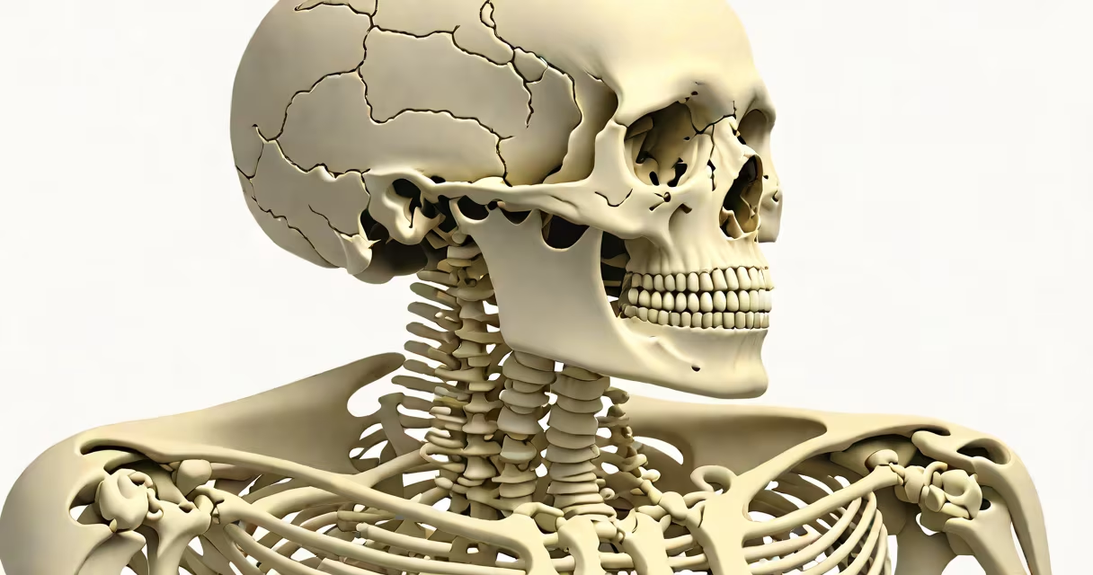
Upper body skeletal anatomy refers to the bones that form the structure of the head, neck, trunk, and limbs above the waist. This system includes the skull, vertebral column, ribcage, shoulder girdle, and arm bones, providing structural support, protecting important organs, and enabling movement.
The upper body skeleton begins at the base of the skull and extends through the cervical, thoracic, and lumbar vertebrae. The cervical spine consists of seven vertebrae (C1-C7), with C1 (atlas) and C2 (axis) forming the joint that allows head rotation. The thoracic spine contains 12 vertebrae, each articulating with a pair of ribs. The lumbar region, though part of the lower back, connects to the upper body via the diaphragm and contributes to trunk stability.
Beneath the neck lies the thoracic cage-a rigid structure formed by the sternum, 12 pairs of ribs, and the thoracic vertebrae. The sternum, or breastbone, is a flat bone that fuses anteriorly and serves as an attachment site for the first seven pairs of ribs (true ribs). The remaining five pairs (false and floating ribs) attach indirectly or not at all to the sternum. This arrangement protects the heart and lungs while allowing controlled expansion during breathing.
The shoulder girdle comprises two bones: the clavicle (collarbone) and the scapula (shoulder blade). The clavicle connects the sternum to the scapula, acting as a strut that maintains shoulder width and absorbs impact. The scapula forms a broad, flat surface against the ribcage and provides attachment points for 17 muscles involved in arm movement.
At the glenohumeral joint, the rounded head of the humerus (upper arm bone) fits into the shallow glenoid cavity of the scapula. This ball-and-socket joint allows many motion, including flexion, extension, abduction, and internal/external rotation. However, the joint's shallow socket makes it inherently unstable-this design prioritizes mobility over stability, which is why shoulder dislocations are common.
The humerus continues down to the elbow, where it articulates with the radius and ulna. The radius is on the thumb side; the ulna is on the little finger side. These bones rotate around each other during pronation and supination-the movements required to turn a doorknob or flip a page.
Each component of the upper body skeleton performs specific mechanical and protective roles. The skull encloses and shields the brain. The vertebral column supports the head and transfers weight to the pelvis. The ribcage protects the heart and lungs, while the sternum stabilizes the front of the chest.
The scapula's acromion process forms the highest point of the shoulder. It articulates with the clavicle at the acromioclavicular (AC) joint. This joint is vulnerable to injury-especially in athletes who fall onto their shoulders. A sprain or dislocation here can cause significant pain and limit overhead motion.
For comparison, the pelvic girdle (lower body) is a closed ring of bones that supports the trunk and transmits forces from the legs to the spine. In contrast, the shoulder girdle is open posteriorly, allowing greater mobility but less stability. This difference explains why the hips are more stable than the shoulders and why shoulder injuries are more frequent in contact sports.
Many assume the ribcage is made of 10 pairs of ribs. In reality, humans have 12 pairs. The first seven pairs (true ribs) connect directly to the sternum. The next three pairs (false ribs) attach to the costal cartilage of the seventh rib. The last two pairs (floating ribs) lack anterior attachments and are free-floating.
Another misconception is that the clavicle is purely decorative. It is not. The clavicle acts as a brace between the sternum and scapula, preventing the shoulder from collapsing inward. It also protects underlying nerves and blood vessels-particularly the brachial plexus and subclavian artery. Fractures of the clavicle are among the most common bone breaks, especially in falls and collisions.
A lesser-known fact: the sternum undergoes ossification throughout life. At birth, it is composed of cartilage. By age 10, it begins to harden. Full fusion of the manubrium, body, and xiphoid process occurs around age 25. This gradual development affects surgical access and trauma response in younger patients.
In a clinical setting, a patient presents with shoulder pain after falling on an outstretched hand. Imaging reveals a fracture of the distal clavicle. The diagnosis hinges on understanding upper body skeletal anatomy: the clavicle's role in shoulder stability, its vulnerability due to location and function, and its relationship to the AC joint.
Without knowledge of the clavicle's anatomy, clinicians might misattribute pain to muscle strain. But recognizing that the clavicle is a critical load-bearing element prevents misdiagnosis. Treatment involves immobilization, physical therapy, and sometimes surgery-depending on displacement and patient activity level.
| Feature | Upper Body | Lower Body |
|---|---|---|
| Bone count (pairs) | 12 pairs of ribs, 2 clavicles, 2 scapulae | 2 hip bones, 2 femurs, 2 tibias, 2 fibulas |
| Joint type (shoulder) | Ball-and-socket (glenohumeral) | Ball-and-socket (hip) |
| Stability vs. mobility | High mobility, moderate stability | High stability, limited mobility |
| Primary function | Manipulation, reach, fine motor control | Weight-bearing, locomotion |
Understanding upper body skeletal anatomy is essential for diagnosing injuries, planning rehabilitation, and appreciating human movement. It underpins fields from orthopedic surgery to physical therapy and sports science.
I once observed a patient with a fractured scapula after a motorcycle crash. The injury was subtle on initial X-ray but became clear on CT scan. The scapular body is thick and dense-fractures often require advanced imaging. This case reinforced how anatomy guides clinical decisions.