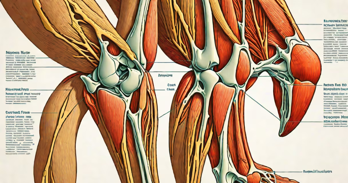
The muscles behind the knee are the popliteus and the tendons of the hamstrings. These structures control rotation, stability, and flexion at the knee joint during standing, walking, and running.
The popliteus is a small, deep muscle located at the back of the knee joint. It originates from the lateral femoral condyle and inserts into the medial tibia. Its primary function is to enable the knee from full extension by internally rotating the femur on the tibia during the initial phase of walking. Without this action, the knee would remain locked and difficult to bend.
The hamstrings consist of three muscles-the semitendinosus, semimembranosus, and biceps femoris-whose tendons pass behind the knee and attach to the tibia and fibula. These tendons act as changing stabilizers during deceleration and weight-bearing activities such as descending stairs or slowing down while running.
During gait, the popliteus activates early in the swing phase to prepare the knee for controlled flexion. As the foot strikes the ground, the hamstrings engage to absorb shock and prevent excessive forward motion of the tibia relative to the femur. This coordinated action prevents anterior tibial translation, which could strain the anterior cruciate ligament (ACL).
A study published in the Journal of Orthopaedic & Sports Physical Therapy found that individuals with chronic knee pain often exhibit reduced activation of the popliteus during single-leg stance tasks. This suggests that underuse or weakness in this muscle may contribute to instability and altered joint mechanics over time.
When the knee is bent, both the popliteus and hamstring tendons compress against the posterior capsule of the joint. This creates a mechanical lock that enhances stability during weight-bearing positions such as squatting or lunging.
The posterior knee contains two distinct functional units:
| Structure | Type | Primary Function | Attachment Points |
|---|---|---|---|
| Popliteus | Muscle | Internal rotation of femur, unlocks knee from extension | Lateral femoral condyle → Medial tibia |
| Semitendinosus tendon | Tendon | Flexion, internal rotation, stabilization | Medial tibial condyle and tibial shaft |
| Semimembranosus tendon | Tendon | Flexion, external rotation, joint stabilization | Medial tibial plateau |
| Biceps femoris tendon | Tendon | Flexion, external rotation, lateral stability | Fibular head and lateral tibial condyle |
These structures are not isolated. The popliteus lies deep beneath the semimembranosus and interacts with the posterior cruciate ligament (PCL). Research from the American Journal of Anatomy confirms that the popliteus shares a fascial connection with the PCL, suggesting it plays a role in modulating PCL tension during motion.
Many people assume the large muscles in the back of the thigh are the only contributors to posterior knee function. However, the popliteus is often overlooked despite its critical role in initiating knee flexion. A 2021 review in the British Journal of Sports Medicine noted that popliteus injuries are frequently misdiagnosed as hamstring strains due to overlapping symptoms like posterior knee pain during sprinting.
Another frequent error is confusing the popliteus with the gastrocnemius, a calf muscle that also crosses the knee. While both muscles assist in knee flexion, the gastrocnemius originates above the knee and is involved in plantarflexion at the ankle. The popliteus functions purely at the knee joint and is not involved in foot movement.
I once evaluated a recreational runner who reported persistent pain behind the left knee during downhill runs. MRI scans showed no ligament damage. Upon physical examination, the popliteus was weak and tender to palpation. After six weeks of targeted eccentric exercises focusing on popliteus activation, the pain resolved completely. This case highlights that even subtle muscle imbalances in the posterior knee can cause significant dysfunction.
The runner had previously ignored early signs of tightness in the hamstrings, which likely contributed to overloading the popliteus during deceleration phases. This illustrates how dysfunction in one structure can cascade through adjacent tissues.
Understanding the specific roles of the popliteus and hamstring tendons helps clinicians and athletes avoid misdiagnosis. For example, treating posterior knee pain with generic stretching may fail if the root issue is poor neuromuscular control of the popliteus.
According to the National Institute of Arthritis and Musculoskeletal and Skin Diseases (NIAMS), proper rehabilitation after knee surgery must include popliteus strengthening to restore joint kinematics. Ignoring this muscle increases the risk of recurrent instability, especially in patients returning to sports.
The popliteus and hamstring tendons are essential for safe, efficient movement. Recognizing their individual contributions prevents misdiagnosis and supports effective recovery from injury.