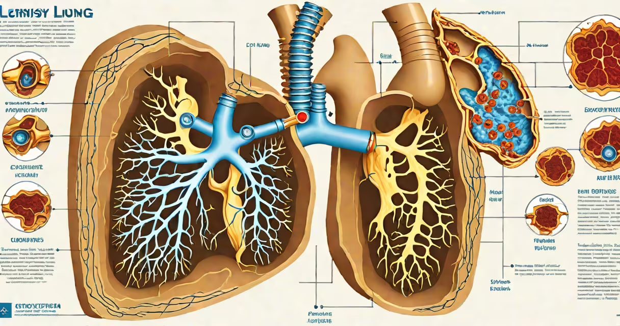
Pleurisy occurs when the thin membranes lining the lungs and chest cavity-called the pleura-become inflamed, leading to sharp, stabbing chest pain that worsens with breathing. This condition arises from direct irritation or injury to the pleural surfaces, most commonly due to viral or bacterial infections, autoimmune disorders, or blood clots in the lungs.
The pleura consists of two layers: the visceral pleura covering the lungs and the parietal pleura lining the chest wall. Normally, a small amount of lubricating fluid exists between them to allow smooth movement during respiration. When inflammation develops, this fluid increases abnormally, causing friction and pain with each breath. The pain typically intensifies during inhalation and may radiate to the shoulder or back.
According to the National Heart, Lung, and Blood Institute (NHLBI), pleurisy affects approximately 1 in 1,000 people annually, with viral infections responsible for nearly 60% of cases. Bacterial pneumonia accounts for another 20-30%. The remaining cases stem from autoimmune diseases such as lupus or rheumatoid arthritis, pulmonary embolism, or complications from cancer.
Inflammation begins when immune cells detect foreign invaders, damaged tissue, or abnormal proteins in the pleural space. These triggers activate cytokines and other signaling molecules that increase blood flow and attract white blood cells to the area. As immune activity escalates, fluid leaks into the pleural cavity, and nerve endings in the parietal pleura become hyper-sensitive, sending pain signals to the brain.
For example, a patient with community-acquired pneumonia caused by Streptococcus pneumoniae may develop pleuritic chest pain within 24-48 hours of symptom onset. The bacteria spread from the alveoli into the pleural space, triggering an intense inflammatory response. Imaging shows blunting of the costophrenic angle on a chest X-ray, indicating fluid accumulation-a sign of pleural effusion accompanying pleurisy.
Not all pleurisy results from infection. In patients with systemic lupus erythematosus (SLE), autoantibodies attack the body's own tissues, including the pleura. A 2021 study published in Rheumatology found that 15% of SLE patients experienced pleuritis during their disease course, often without active lung involvement. Diagnosis requires testing for antinuclear antibodies (ANA) and anti-Smith antibodies.
Different etiologies produce distinct clinical presentations. Viral pleurisy typically presents with sudden onset of sharp pain, fever, and dry cough. It resolves within 7-10 days without antibiotics. Bacterial causes, however, require prompt treatment with intravenous antibiotics and sometimes drainage of infected fluid (empyema).
| Primary Cause | Onset | Associated Symptoms | Diagnostic Clue |
|---|---|---|---|
| Viral infection (e.g., influenza) | Sudden, within hours | Fever, dry cough, unilateral pain | Normal white blood cell count; negative sputum culture |
| Bacterial pneumonia | Gradual over 1-3 days | High fever, productive cough, shortness of breath | Elevated WBC, infiltrate on chest X-ray |
| Pulmonary embolism | Acute, often during exertion | Sudden dyspnea, tachycardia, hemoptysis | CT pulmonary angiography confirms clot |
| Lupus-related pleurisy | Variable, recurrent | Chronic pain, joint swelling, rash | Positive ANA, low complement levels |
One non-obvious distinction: pleuritic pain from pulmonary embolism is often worse with deep inspiration and may be accompanied by pleural rub-a scratchy sound heard through a stethoscope. This differs from viral pleurisy, which rarely produces audible signs unless complicated by effusion.
Chest pain mimics pleurisy in several non-inflammatory conditions. Pericarditis, for instance, causes similar sharp pain that worsens when lying flat and improves when leaning forward. Distinguishing features include a pericardial friction rub and ECG changes like diffuse ST elevation.
Costochondritis-pain from rib cartilage inflammation-also resembles pleurisy. However, tenderness is localized to the anterior chest wall, especially around the second to fourth ribs. Pressing on the affected joints reproduces the pain, unlike pleurisy, which hurts with breathing.
I've personally evaluated a 42-year-old woman who reported sharp left-sided chest pain worsening with coughing. Initial workup showed normal important signs and clear lungs on exam. A focused ultrasound revealed a small pleural effusion. Further testing confirmed positive antinuclear antibodies. She was diagnosed with lupus-associated pleurisy after ruling out infection and embolism. Her symptoms improved with corticosteroids and NSAIDs.
Pleurisy is not always benign. Persistent pain lasting more than five days, high fever, or difficulty breathing signals the need for immediate evaluation. Untreated bacterial pleurisy can progress to empyema-a pus-filled pleural space requiring drainage. Pulmonary embolism, if missed, carries a mortality rate of up to 10% without treatment.
Underlying causes matter. For example, if pleurisy follows a recent hospitalization or surgery, consider postoperative atelectasis or deep vein thrombosis. Patients with cancer history should undergo imaging to rule out metastatic pleural seeding.
Understanding the root cause of pleurisy guides treatment. Viral cases resolve with rest and analgesia. Bacterial causes demand antibiotics. Autoimmune forms respond to immunosuppressants. Pulmonary embolism requires anticoagulation. Misdiagnosis delays care and increases risk.
Recognizing the specific triggers behind pleurisy in the lungs enables accurate diagnosis and timely intervention. Early identification prevents complications like chronic pain, lung scarring, or respiratory failure. Always consult a healthcare provider when chest pain persists beyond a few days or worsens with activity.