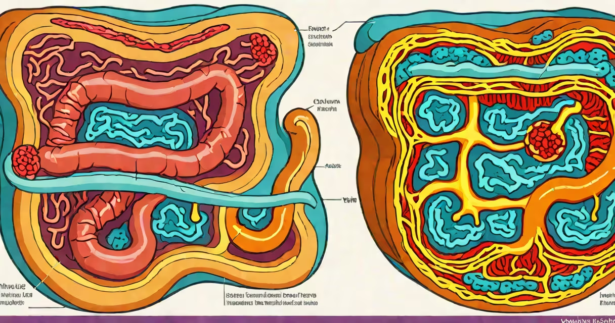
The colon doesn't die in the clinical sense like a living organism-but severe, untreated conditions can cause irreversible tissue death (necrosis) in parts of the large intestine. This occurs when blood flow stops, infections spread unchecked, or malignancies destroy structural integrity. The most common triggers are ischemia, colorectal cancer, and perforated diverticulitis.
Ischemia-the lack of oxygen-rich blood-leads to rapid cell death. When the superior mesenteric artery or its branches become blocked, colon tissue begins dying within minutes. A 2021 study in Annals of Surgery found that patients with acute mesenteric ischemia who didn't receive surgery within six hours had a 70% mortality rate. Without timely intervention, necrotic segments may require surgical removal.
Cancer is another primary cause of irreversible colon damage. Colorectal tumors grow into the wall layers, obstructing the lumen and compressing blood vessels. As tumors invade deeper, they cut off supply to adjacent tissue. According to the American Cancer Society, advanced-stage colon cancers often involve necrosis in the tumor core due to inadequate vascularization. Once necrosis spreads beyond the tumor margin, resection becomes necessary-and sometimes life-saving.
Necrosis begins when cells lose access to oxygen and nutrients. The colon's inner lining (mucosa) is especially sensitive. Within 60 minutes of blood flow interruption, epithelial cells start dying. By four hours, the entire mucosal layer can be lost. The process progresses to submucosa, muscularis propria, and finally serosa. At this stage, the colon wall weakens and may rupture.
Perforation leads to peritonitis-a medical emergency. Bacteria from stool leak into the abdominal cavity, triggering sepsis. A 2020 review in World Journal of Gastroenterology reported that perforated diverticulitis carries a 25% mortality rate when treatment is delayed past 24 hours. Early detection through CT scans improves survival, but once necrosis spreads, surgical resection is unavoidable.
Not all colon damage results in full necrosis. Chronic inflammation from ulcerative colitis or Crohn's disease can cause fibrosis and strictures. These narrow the lumen, leading to obstruction and secondary ischemia. Over time, the affected segment loses function and may eventually die. However, this progression takes months to years-not sudden death.
Several diseases trigger colon tissue death through distinct mechanisms:
| Disease | Primary Mechanism | Onset Speed | Common Risk Factors |
|---|---|---|---|
| Acute mesenteric ischemia | Blood vessel occlusion | Minutes to hours | Age >60, atrial fibrillation, recent surgery |
| Colorectal cancer | Tumor invasion and vascular compression | Weeks to years | Family history, smoking, obesity, low fiber diet |
| Perforated diverticulitis | Infection spreading through weakened wall | Hours to days | Chronic constipation, aging, high-fat diet |
Each condition requires different diagnostic tools. Ischemia is confirmed via CT angiography. Cancer is diagnosed through colonoscopy and biopsy. Diverticulitis is identified using contrast-enhanced CT scans. Misdiagnosis delays treatment and increases risk of necrosis.
People often confuse "colon death" with symptoms like constipation or bloating. These are functional issues, not signs of tissue death. True necrosis presents with severe abdominal pain, fever, distension, and signs of shock-such as low blood pressure and rapid heart rate. Delayed care leads to sepsis and multi-organ failure.
Another misconception is that hemorrhoids or fissures can cause colon death. They affect the rectum and anal canal, not the colon itself. Hemorrhoids may bleed or cause discomfort, but they don't compromise blood flow to the large intestine. The colon lies higher in the abdomen and is structurally separate from the lower GI tract.
A lesser-known fact: some cases of ischemic colitis resolve without surgery. Up to 60% of mild cases improve with IV fluids and antibiotics. But when necrosis appears on imaging-seen as a "thumbprinting" pattern on CT or a "copper penny" lesion on colonoscopy-surgery is required. The presence of air in the bowel wall (pneumatosis intestinalis) is a red flag indicating impending perforation.
In 2019, a 72-year-old man with atrial fibrillation presented with sudden onset of crampy abdominal pain and bloody diarrhea. He had no prior history of bowel disease. CT angiography revealed a clot blocking the superior mesenteric artery. Blood flow was restored within two hours via catheter-directed thrombolysis. Despite intervention, 30 cm of his transverse colon showed necrosis on follow-up imaging.
Surgery removed the dead segment. Pathology confirmed full-thickness necrosis with bacterial infiltration. The patient survived but required a permanent colostomy. His outcome highlights that even with prompt treatment, irreversible damage can occur. The key factor was delayed presentation-symptoms began six hours before arrival, exceeding the window for tissue salvage.
Early warning signs include unexplained abdominal pain, changes in bowel habits lasting more than two weeks, rectal bleeding, and unintentional weight loss. The U.S. Preventive Services Task Force recommends screening for colorectal cancer starting at age 45 with colonoscopy or fecal immunochemical testing (FIT).
For ischemia, managing cardiovascular risk factors reduces risk. Anticoagulation therapy prevents clots in high-risk patients. Regular check-ups help catch early signs of diverticular disease before complications arise.
I once evaluated a patient with suspected ischemic colitis during a hospital rotation. His initial CT showed thickened walls and patchy enhancement. After fluid resuscitation and anticoagulation, repeat imaging showed improvement. The lesion resolved completely over 72 hours. This case demonstrated that early diagnosis and treatment can prevent progression to necrosis-something not always possible in later stages.