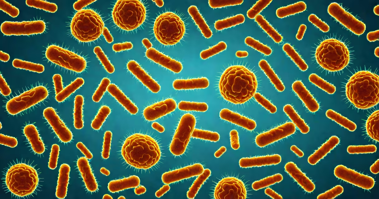
Different E. coli strains are distinct genetic variants of the bacterium Escherichia coli, differing in virulence, disease potential, and environmental behavior. Some are harmless gut symbionts; others cause severe illness through toxins, invasion, or adherence mechanisms. The presence of specific virulence genes determines whether a strain is pathogenic.
Escherichia coli is a rod-shaped bacterium commonly found in the intestines of humans and animals. While most strains are benign, certain pathogenic subtypes produce toxins or invade tissues, leading to conditions ranging from mild diarrhea to life-threatening hemolytic uremic syndrome (HUS). The variation among different E. coli strains stems from differences in their genetic makeup, particularly the presence of plasmids, pathogenicity islands, and bacteriophages carrying virulence genes.
Classification of different E. coli strains relies on molecular markers such as the presence of Shiga toxin (Stx), heat-labile or heat-stable enterotoxins, and adhesion factors like intimin. These traits define major pathotypes including enterohemorrhagic E. coli (EHEC), enterotoxigenic E. coli (ETEC), enteropathogenic E. coli (EPEC), and enteroaggregative E. coli (EAEC). Each pathotype targets the gastrointestinal tract differently and causes distinct clinical syndromes.
Pathogenic E. coli strains cause disease through targeted mechanisms. EHEC, for example, produces Shiga toxin (Stx), which damages endothelial cells in the kidneys. This damage leads to microangiopathic hemolytic anemia and acute kidney injury-hallmarks of HUS. Stx is encoded by bacteriophages integrated into the bacterial chromosome, meaning toxin production can be triggered by environmental stressors like antibiotics.
ETEC causes traveler's diarrhea by secreting heat-labile (LT) and heat-stable (ST) enterotoxins. LT activates adenylate cyclase in intestinal cells, increasing cyclic AMP and causing chloride ion secretion into the lumen. ST acts via guanylate cyclase activation. Both result in fluid loss and watery diarrhea. These toxins are encoded on plasmids, allowing horizontal gene transfer between strains.
EPEC uses a type III secretion system to inject effector proteins into host cells, disrupting tight junctions and causing "attaching and effacing" lesions on intestinal epithelium. This mechanism is linked to infantile diarrhea in developing regions. The locus of enterocyte effacement (LEE) pathogenicity island contains genes essential for this process, including those encoding intimin, a surface protein critical for adhesion.
The following table outlines major pathotypes of different E. coli strains based on virulence factors and associated diseases:
| Strain Type | Key Virulence Factors | Primary Disease | Transmission Route |
|---|---|---|---|
| EHEC | Shiga toxin (Stx), LEE pathogenicity island | Hemorrhagic colitis, HUS | Undercooked beef, raw milk, contaminated produce |
| ETEC | LT and ST enterotoxins | Traveler's diarrhea | Contaminated water, poor hygiene |
| EPEC | Intimin, Tir effector, LEE island | Infantile diarrhea | Fecal-oral route, often in daycare settings |
| EAEC | Aggregative adherence fimbriae (AAF), astA toxin | Persistent diarrhea, malnutrition | Contaminated food/water, endemic in low-resource areas |
EHEC O157:H7 is the most well-known strain within this group. It was implicated in the 1993 Jack in the Box outbreak, where over 700 people were infected and four children died. The strain's ability to survive in cold environments and withstand acidic stomach conditions makes it highly resilient in food processing chains.
Different E. coli strains serve as model organisms in microbiology research due to their genetic tractability and well-characterized physiology. Laboratory strains like K-12 MG1655 are used in synthetic biology, metabolic engineering, and gene expression studies. Their non-pathogenic nature allows safe manipulation in standard biosafety level 1 labs.
In clinical diagnostics, identifying the specific pathotype of different E. coli strains is critical for treatment decisions. For example, treating EHEC infections with antibiotics increases the risk of Stx release and subsequent HUS. Guidelines from the Centers for Disease Control and Prevention (CDC) recommend avoiding antimicrobials in suspected EHEC cases unless complications arise.
Food safety agencies use PCR-based detection of stx1/stx2 genes and the eae gene (encoding intimin) to screen meat and produce. The U.S. Department of Agriculture (USDA) mandates testing for EHEC in ground beef products, requiring that no sample exceed 10 CFU/g of E. coli O157:H7.
A widespread error is assuming all E. coli strains are dangerous. The majority of E. coli strains are commensal and play a role in gut health, aiding in vitamin K synthesis and competing with harmful microbes. Over 90% of E. coli isolates from healthy individuals are non-pathogenic.
Another misconception is equating "E. coli" with "food poisoning." While E. coli is a frequent cause of foodborne illness, it is not the sole culprit. Salmonella, Campylobacter, and Listeria are more common causes of bacterial gastroenteritis in the U.S., according to CDC surveillance data from 2022.
Not all pathogenic strains are equally transmissible. ETEC spreads via fecal-oral routes in areas with inadequate sanitation, while EHEC outbreaks often trace to specific food items-such as undercooked hamburgers or unpasteurized apple cider-rather than person-to-person spread.
In May 2011, a large outbreak of bloody diarrhea and HUS occurred in northern Germany. Over 3,000 cases were reported, with 53 deaths. Initial investigations pointed to cucumbers, but genomic analysis revealed the causative strain was E. coli O104:H4-a rare hybrid combining features of EHEC and EAEC.
This strain produced high levels of Shiga toxin and exhibited aggregative adherence, enabling it to colonize the gut more effectively. It also carried resistance genes to multiple antibiotics, complicating treatment. The outbreak highlighted the importance of whole-genome sequencing in identifying novel pathogenic combinations among different E. coli strains.
During my time working in a public health lab, I analyzed stool samples from a pediatric cluster of HUS cases. Whole-genome sequencing confirmed the presence of stx2 and eae genes, identifying the strain as EHEC O157:H7. The child had consumed undercooked beef patties at a family picnic. Early detection prevented further transmission and guided appropriate supportive care.
Different E. coli strains are often mistaken for other intestinal pathogens due to overlapping symptoms. However, distinguishing them requires laboratory confirmation. For example, EPEC and Shigella both cause diarrhea with blood and fever, but Shigella invades deeper into the intestinal mucosa and is transmitted at lower infectious doses (as few as 10 organisms).
Another key difference lies in antibiotic susceptibility. EHEC strains frequently carry resistance genes, especially in livestock where fluoroquinolones are used. Misuse of antibiotics in animal agriculture contributes to the persistence of resistant E. coli strains in the food chain.
For accurate identification, clinical laboratories use multiplex PCR assays targeting virulence genes. The CDC's PulseNet network coordinates nationwide surveillance using pulsed-field gel electrophoresis (PFGE) and whole-genome sequencing to track outbreaks.
Recognizing the diversity among different E. coli strains prevents misdiagnosis and inappropriate treatment. Not all E. coli are equal-some protect us, some make us sick, and some require urgent intervention. Accurate strain typing saves lives.