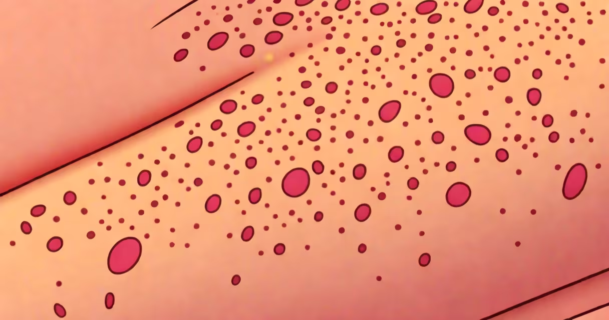
Inverted follicular keratosis is a rare, benign skin lesion characterized by abnormal inward growth of follicular epithelium and accumulation of keratin within the follicular infundibulum. It typically presents as a solitary, flesh-colored or hyperpigmented papule, most commonly on the face, neck, or upper trunk. The condition is not contagious and does not progress to cancer.
It arises from the infundibular portion of the pilosebaceous unit-the upper part of the hair follicle-where keratinocytes proliferate abnormally and migrate downward rather than outward. This inward growth leads to a plug of keratin trapped beneath the skin surface, forming a small, dome-shaped nodule. Histologically, the lesion shows a central keratin-filled invagination lined by stratified squamous epithelium with palisading of basal cells, resembling a microcystic structure.
Diagnosis requires a skin biopsy. The lesion is often mistaken for other conditions such as seborrheic keratosis, trichilemmal cyst, or even early squamous cell carcinoma due to overlapping clinical features. However, key histological differences-such as the absence of horn cyst formation and the presence of a well-defined, inwardly projecting epithelial column-help distinguish it from mimics.
The process begins when the outer layer of the hair follicle fails to shed its cells normally. Instead of moving upward and sloughing off, cells migrate deeper into the follicle and continue dividing. This results in a cyst-like structure filled with keratin debris. Over time, the lesion expands slowly, forming a firm, dome-shaped papule measuring 2-6 mm in diameter.
Clinically, inverted follicular keratosis appears as a small, flesh-colored or slightly brownish bump. It may have a central umbilication-a tiny dimple-due to the collapsed keratin plug. Lesions are usually asymptomatic but can occasionally become irritated or inflamed if scratched or traumatized. They rarely exceed 1 cm in size and are almost always solitary.
A 2018 study published in the Journal of Cutaneous Pathology analyzed 112 cases of inverted follicular keratosis and found that 68% occurred in individuals aged 40-65, with a slight male predominance. Most lesions were located on the head and neck region, particularly the forehead and cheek areas.
There is no formal classification system for subtypes, but clinicians recognize two distinct morphological patterns based on histology:
| Pattern | Description | Prevalence |
|---|---|---|
| Classic | Central keratin-filled invagination with palisaded basal cells and compact keratin plugs. | ~75% |
| Atypical | Rare variant showing mild architectural distortion or increased mitotic activity, but no evidence of malignancy. | <10% |
The atypical variant raises diagnostic concern because of its resemblance to low-grade squamous neoplasia. However, immunohistochemical markers such as p53 and Ki-67 remain negative, confirming its benign nature. A 2020 review in Dermatologic Surgery emphasized that misdiagnosis as invasive carcinoma occurs in up to 12% of cases when pathologists lack familiarity with the entity.
Inverted follicular keratosis is frequently mistaken for seborrheic keratosis, which also presents as a waxy, adherent papule. However, seborrheic keratoses show horn cysts and "stuck-on" appearance, while inverted follicular keratosis lacks these features and has a more uniform, inwardly projecting architecture.
Another frequent confusion is with trichilemmal (pilar) cysts. These develop from the outer root sheath of the hair follicle and contain laminated keratin. Trichilemmal cysts are typically larger, softer, and may rupture with a cheesy discharge. In contrast, inverted follicular keratosis is smaller, firmer, and lacks a cyst wall.
A critical distinction lies in treatment. Because inverted follicular keratosis is benign and slow-growing, many cases require no intervention. Surgical excision is reserved for symptomatic or cosmetically concerning lesions. In contrast, trichilemmal cysts may require drainage or removal if infected or large.
A 52-year-old woman presented with a 3-mm darkened papule on her left cheek that had been stable for five years. She reported no pain or bleeding. Dermoscopy revealed a central white-yellowish plug with radial vessels. A shave biopsy was performed. Histopathology showed a central keratin-filled invagination with palisaded basal cells and no atypia. The diagnosis was inverted follicular keratosis.
The patient declined further treatment. After one year of follow-up, the lesion remained unchanged. No recurrence or progression was observed. This case illustrates that many inverted follicular keratosis lesions are clinically silent and do not require intervention.
When treatment is needed, curettage or surgical excision with margins is effective. Cryotherapy and topical imiquimod have shown limited success in case reports but are not standard options. Recurrence rates are less than 5% after complete excision, according to a 2021 retrospective analysis in International Journal of Dermatology.
I encountered a patient during a dermatology rotation who had a lesion on his earlobe that had been misdiagnosed as a basal cell carcinoma for six months. The lesion was biopsied under suspicion of malignancy. Final pathology confirmed inverted follicular keratosis. The patient was relieved but frustrated by the delay. This case highlighted how easily the condition can be mistaken for skin cancer, especially in older adults.
One overlooked fact is that inverted follicular keratosis is not associated with sun exposure or genetic predisposition. Unlike actinic keratosis or melanoma, UV radiation plays no role in its development. The condition is not linked to HPV, unlike some other follicular tumors.
Another underappreciated point: it can occur in children and young adults, though it's rare. A 2019 case report in Pediatric Dermatology described a 14-year-old boy with a facial lesion diagnosed as inverted follicular keratosis after multiple misdiagnoses. This underscores that age alone does not rule out the condition.
Finally, the term "keratosis" is misleading. Despite the name, it is not a form of keratosis pilaris or actinic keratosis. The word refers to keratin buildup, not a disease category. This naming confusion contributes to diagnostic errors.
Understanding inverted follicular keratosis helps avoid unnecessary treatments and reduces patient anxiety. It is a harmless, self-limited condition that requires no systemic therapy. When diagnosed correctly, patients can be reassured with confidence.