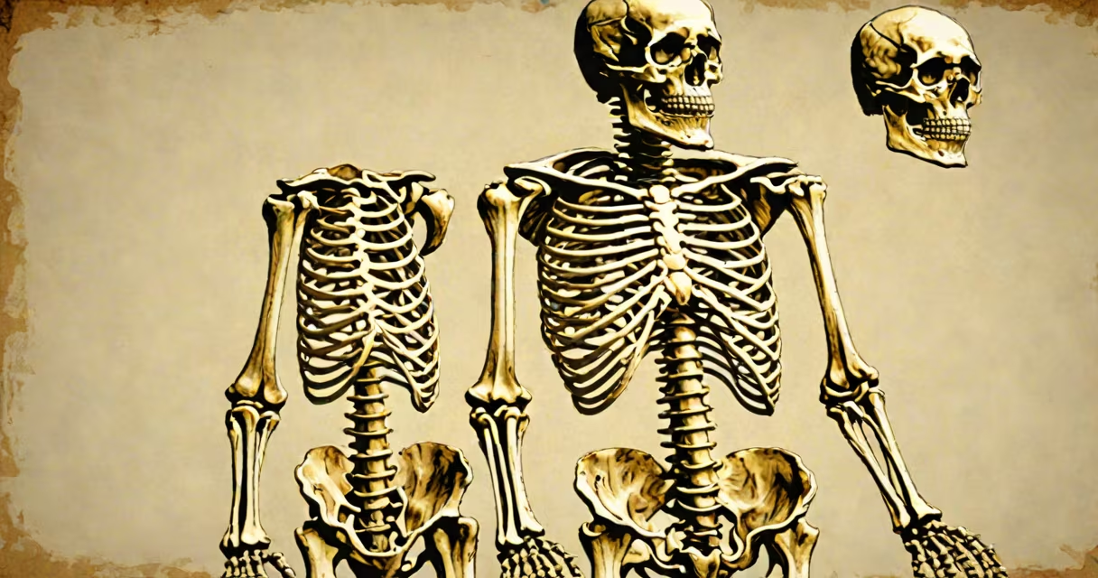
A skeleton life size model is a full-scale, anatomically accurate replica of the human skeleton, designed for educational, medical, and display purposes. It replicates the 206 bones of the adult human body in precise proportions and spatial relationships.
These models are typically constructed from durable materials such as PVC, resin, or polyethylene, with joints that allow for articulation. They are used in classrooms, medical schools, museums, and clinical training environments to teach bone structure, joint mechanics, and human posture.
Unlike scaled-down or simplified versions, a life size model maintains the actual dimensions of the human skeleton-approximately 1.7 meters (5'7") tall for an average adult. This scale enables learners to grasp spatial relationships between bones, understand weight distribution, and visualize movement patterns in real-world context.
The functionality of a skeleton life size model lies in its structural fidelity. Each bone is molded to match the real human counterpart-femur, scapula, vertebrae, and cranial bones-all positioned according to anatomical standards. The spine, for example, includes seven cervical, twelve thoracic, and five lumbar vertebrae, with correct curvature and alignment.
Articulated models feature movable joints at the hips, shoulders, elbows, and knees. These allow instructors to demonstrate range of motion, dislocation, and biomechanical strain. Some models include removable parts-such as the skull cap or pelvis-for hands-on study of internal structures.
For example, a model used in a university anatomy lab may have labeled bones and color-coded ligaments. When a student rotates the shoulder joint, they observe how the humerus interacts with the glenoid cavity, reinforcing concepts taught in textbooks.
Skeleton life size models vary by material, level of detail, and intended use. The most common types are:
| Type | Material | Use Case | Key Feature |
|---|---|---|---|
| Standard Plastic | PVC or polyethylene | Classroom instruction | Cost-effective, lightweight, durable |
| Articulated Metal | Aluminum frame with plastic bones | Medical training, surgery prep | Adjustable joints, realistic resistance |
| High-Fidelity Resin | Cast resin with surface texture | Museum displays, research | Textured bone surfaces, lifelike finish |
| Dissectible Model | Composite materials with removable components | Advanced anatomy courses | Removable skull, rib cage, or pelvic bones |
Each type serves different needs. A standard plastic model costs $150-$300 and is ideal for high school biology classes. In contrast, a high-fidelity resin model can cost $1,200 or more and is used in university labs for detailed study of bone density and fracture patterns.
Life size skeleton models are essential tools in health sciences education. According to the American Association of Anatomists, 87% of U.S. medical schools use full-size anatomical models during gross anatomy instruction. These models reduce reliance on cadavers, which are limited in supply and require specialized storage.
In physical therapy programs, models help trainees assess gait abnormalities. For instance, a therapist studying a patient with a limp uses a skeleton life size model to simulate altered hip rotation and observe how it affects knee stress. This allows for early intervention planning.
Museums also use these models to engage the public. The Smithsonian National Museum of Natural History features a full-size skeleton model in its Human Origins exhibit. It displays the evolution of bipedalism through changes in the pelvis and femur angles across species- sapiens.
Many assume that all skeleton models are identical. In reality, accuracy varies widely. Some low-cost models distort proportions-such as elongating the femur or misaligning the sacrum. These errors lead to incorrect understanding of balance and movement.
Another misconception is that a skeleton life size model replaces cadaver dissection. While useful, it cannot replicate tissue elasticity, blood vessel networks, or nerve pathways. The National Institutes of Health notes that tactile experience with real tissue remains irreplaceable for surgical training.
A key distinction: a skeleton life size model is not a mannequin. Mannequins are full-body figures with skin, muscle, and organs. A skeleton model shows only bones and joint connections. Confusing the two leads to flawed anatomical reasoning.
I once observed a medical student using a skeleton life size model during a musculoskeletal exam rehearsal. She adjusted the pelvis to simulate anterior tilt and noted how it shifted the center of gravity. The model's articulated hip joints allowed her to feel resistance, mimicking real joint stiffness. This hands-on practice helped her diagnose a patient's gait issue more accurately during clinical rotations.
Most skeleton life size models are not cast from real human skeletons. Instead, they are based on standardized anatomical references such as the Frankfort horizontal plane and the International Federation of Associations of Anatomists (IFAA) guidelines. This ensures consistency across institutions-even when models are manufactured in different countries.
For example, a model produced in China and another in Germany both follow the same reference points for the position of the mandible relative to the occipital condyles. This standardization prevents confusion in global medical education.