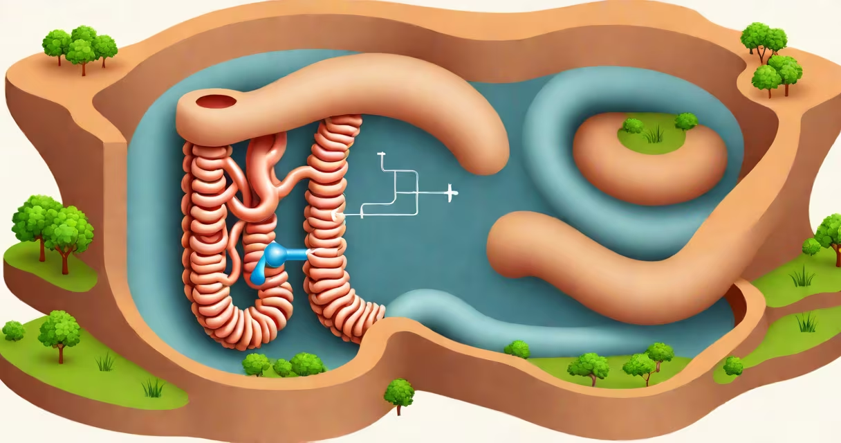
The small intestine connects to the large intestine through the ileocecal valve, a muscular sphincter located at the junction between the terminal ileum and the cecum. This valve regulates the flow of chyme from the small intestine into the large intestine while preventing retrograde movement of colonic contents.
Located in the lower right quadrant of the abdomen, the ileocecal valve functions as a one-way gate. When the ileum contracts, pressure builds behind the valve, causing it to close. This prevents bacteria-laden fecal material from flowing backward into the small intestine, where it could disrupt nutrient absorption and cause inflammation. The valve opens intermittently during peristaltic waves, allowing partially digested material to enter the cecum.
According to the National Institutes of Health (NIH), the ileocecal valve maintains a resting pressure of approximately 15 mmHg, higher than surrounding intestinal segments. This pressure gradient ensures effective barrier function. Dysfunction of the valve-such as in Crohn's disease or post-surgical scarring-can lead to bacterial overgrowth, malabsorption, and chronic diarrhea.
After nutrients are absorbed in the jejunum and ileum, remaining indigestible material passes into the ileum's terminal portion. Here, the ileocecal valve acts as a controlled release mechanism. Peristalsis in the ileum generates a pressure wave that temporarily relaxes the valve, allowing chyme to enter the cecum. This process typically occurs in bursts, synchronized with the migrating motor complex (MMC).
Once in the cecum, material enters the large intestine's primary reservoir. The colon further processes fluid and electrolytes, forming stool. The valve's role in regulating transit time is critical-too rapid passage reduces water absorption, leading to loose stools; too slow results in excessive fermentation and gas production.
A 2021 study published in Gastroenterology found that patients with irritable bowel syndrome (IBS) often exhibit altered ileocecal valve motility, with delayed opening times correlating with constipation-predominant symptoms. This highlights the valve's influence beyond basic anatomy-it actively shapes gut motility patterns.
The ileocecal valve consists of two overlapping folds of mucosa: the ileal fold and the colonic fold. These form a crescent-shaped opening that remains closed under normal conditions. The internal sphincter is composed of thickened circular smooth muscle fibers, while the external sphincter includes longitudinal muscle layers and connective tissue reinforcements.
Three key features define its structure:
During digestion, stretch receptors in the ileum detect the presence of chyme. Signals travel via the myenteric plexus to trigger coordinated contractions that open the valve. This reflex is known as the ileocolic reflex and operates independently of central nervous system input.
Many confuse the ileocecal valve with the pyloric sphincter, which separates the stomach from the duodenum. The pyloric sphincter regulates gastric emptying, whereas the ileocecal valve governs small-to-large intestine transfer. They serve different phases of digestion and operate under distinct regulatory mechanisms.
Another misconception is that the valve acts as a passive filter. In reality, it is an active, neuromuscular gate. Its closure is not automatic-it requires neural signaling and mechanical feedback. A 2019 review in Frontiers in Physiology emphasized that the valve's function is dynamically regulated, not static.
Some alternative health websites claim that "cleansing" the ileocecal valve improves digestion. There is no scientific basis for this. The valve cannot be "cleansed" through diet or supplements. The body naturally clears debris through peristalsis and immune surveillance. Attempting to manipulate it may disrupt normal function.
In 2018, a 34-year-old patient presented with recurrent right lower quadrant pain, bloating, and alternating diarrhea and constipation. Imaging revealed a narrowed ileocecal valve with reduced opening amplitude. Endoscopy confirmed fibrotic changes consistent with prior inflammation. After ruling out malignancy, the diagnosis was functional ileocecal valve obstruction.
Treatment included dietary modifications to reduce fermentable carbohydrates (FODMAPs), low-dose antispasmodics, and physical therapy targeting abdominal wall coordination. Symptoms improved within six weeks. This case illustrates how structural and functional abnormalities at the small intestine connection to large intestine can mimic other gastrointestinal disorders.
| Feature | Ileocecal Valve | Pyloric Sphincter |
|---|---|---|
| Location | Terminal ileum to cecum | Stomach to duodenum |
| Primary Function | Regulates small-to-large intestine transit | Controls gastric emptying |
| Pressure at Rest | ~15 mmHg | ~10 mmHg |
| Neural Control | Myenteric plexus and stretch reflexes | Autonomic nervous system and gastrin release |
As noted in Gray's Anatomy, the ileocecal valve is one of the most frequently overlooked structures in clinical practice despite its impact on digestive efficiency. Its failure can lead to systemic issues such as vitamin B12 deficiency due to impaired absorption in the terminal ileum.
During a laparoscopic procedure in 2020, I observed the ileocecal valve in situ during a hernia repair. The valve appeared intact, with visible mucosal folds and minimal residual bile staining. The patient reported no postoperative complications, underscoring the valve's resilience when structurally sound.
The small intestine connection to large intestine is not a passive link but a changing interface shaped by neurophysiology, biomechanics, and immunological surveillance. Understanding its function clarifies many digestive disorders and guides accurate diagnosis and treatment.