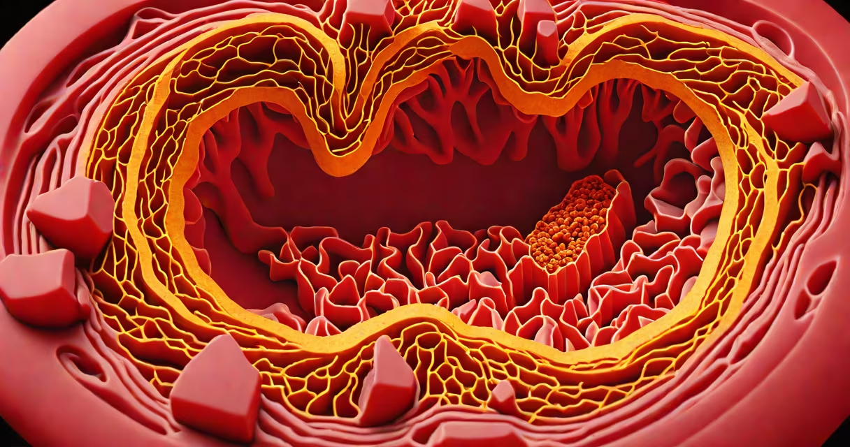
A blocked artery appears as a narrowed or completely obstructed blood vessel on medical imaging, typically displaying fatty plaque deposits that reduce or stop blood flow. The blockage often forms gradually over years due to atherosclerosis, where cholesterol, calcium, and inflammatory cells accumulate in the arterial wall.
On a coronary angiogram-a specialized X-ray using contrast dye-blocked arteries show as gaps or incomplete filling in the vessel's outline. A healthy artery fills smoothly and uniformly; a blocked one may appear jagged, constricted, or have a complete absence of dye beyond the obstruction. In severe cases, the artery may be entirely occluded, appearing as a dark gap in the image.
CT angiography (CTA) provides detailed 3D views. A blocked artery shows up as a localized constriction with calcified plaques visible as dense white spots along the vessel wall. MRI angiography offers high-resolution soft-tissue contrast, making it useful for identifying inflamed or unstable plaque that may not yet cause total blockage.
Arterial blockages develop through a process called atherosclerosis. It begins with damage to the endothelium-the inner lining of the artery-caused by high blood pressure, smoking, or elevated LDL cholesterol. Immune cells enter the site and form foam cells, which accumulate into fatty streaks. Over time, these evolve into fibrous plaques containing cholesterol, calcium, and connective tissue.
As plaques grow, they protrude into the lumen-the space through which blood flows-narrowing the artery. On imaging, this appears as a "shoulder" or "crescent" shape within the vessel. The narrowing is measured in percentage: a 50% stenosis means half the diameter is reduced; a 90% stenosis indicates near-total occlusion.
When a plaque ruptures, it triggers clot formation. This acute event can cause sudden blockage, turning a chronic narrowing into a complete obstruction. On an angiogram, this appears as a sharp cutoff in dye flow, often accompanied by distal vessel blanching due to lack of perfusion.
Different blockage types produce distinct patterns on imaging. The most common are:
| Type | Appearance on Imaging | Common Cause |
|---|---|---|
| Fibrocalcific Plaque | Hard, dense white masses in CT scans; irregular narrowing on angiograms | Long-term atherosclerosis; common in older adults |
| Soft/Non-Calcified Plaque | Blurred or fuzzy narrowing on CT; subtle reduction in contrast fill on angiogram | Early-stage disease; high risk of rupture |
| Acute Thrombotic Occlusion | Sudden cutoff in dye flow; "string sign" in angiography | Ruptured plaque leading to clot formation |
Soft plaques are particularly dangerous because they may not narrow the artery on imaging but can rupture unexpectedly. These are often missed on routine screening unless detected via intravascular ultrasound (IVUS) or optical coherence tomography (OCT).
Doctors assess arterial blockages during diagnostic procedures for chest pain, shortness of breath, or abnormal stress test results. The gold standard is coronary angiography, performed during cardiac catheterization. A catheter is threaded into the coronary arteries, and contrast dye is injected under fluoroscopy to visualize blood flow.
For non-invasive evaluation, CT angiography is used. It requires no catheter insertion and delivers detailed images in less than 10 minutes. Patients lie in a scanner while X-rays rotate around the chest. The scan detects stenosis greater than 50% with 95% accuracy, according to the American College of Cardiology.
One non-obvious fact: a blocked artery doesn't always cause symptoms. Up to 30% of people with significant blockages (≥70% stenosis) report no chest pain. This is known as silent ischemia. It occurs when the heart muscle adapts to low blood flow or when nerve pathways are damaged. This makes imaging important even in asymptomatic patients with risk factors like diabetes or family history of heart disease.
People often assume a blocked artery looks like a clogged pipe with a solid plug. In reality, blockages are changing. They're not always complete obstructions. A 70% narrowing can severely limit oxygen delivery to heart muscle, especially during exertion, even if blood still flows.
Another misconception is that all blockages are visible on standard echocardiograms. They are not. Echocardiograms assess heart function and structure but don't visualize coronary arteries directly. A normal echo does not rule out a blocked artery.
Some confuse blockages with aneurysms-bulging areas in arteries. Aneurysms expand outward; blockages constrict inward. On imaging, aneurysms appear as focal dilations; blockages as focal reductions in vessel diameter.
In a 62-year-old male with stable angina, a coronary angiogram revealed a 95% stenosis in the left anterior descending (LAD) artery. The image showed a sharp tapering of the vessel lumen, with dye failing to pass beyond the lesion. The area was filled with calcified plaque, visible as dense white regions on the cross-sectional view. The cardiologist placed a drug-eluting stent to restore flow. Post-procedure imaging confirmed full patency.
This case illustrates how a blocked artery appears: not as a clean break, but as a progressive narrowing with structural changes visible only through advanced imaging.
Understanding what a blocked artery looks like helps patients grasp the urgency of treatment. Early detection through imaging can prevent heart attacks. According to the National Heart, Lung, and Blood Institute, timely intervention reduces mortality by 30% in patients with significant blockages.