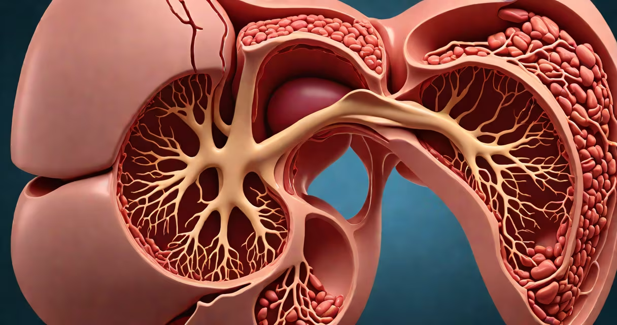
Enlarged liver and spleen-also called hepatosplenomegaly-indicate that both organs are larger than normal, often signaling an underlying condition such as infection, liver disease, or blood disorder. This finding is typically detected via imaging like ultrasound or CT scan during evaluation for fatigue, abdominal pain, or unexplained weight loss.
When the liver and spleen are both enlarged, it suggests systemic involvement rather than isolated organ dysfunction. The liver filters toxins and produces proteins; the spleen removes old blood cells and supports immune function. Their simultaneous enlargement points to a process affecting the bloodstream or internal organs broadly.
For example, a 45-year-old woman with persistent fatigue and mild jaundice had an ultrasound revealing a liver measuring 18 cm (normal: 12-15 cm) and a spleen measuring 16 cm (normal: 8-12 cm). Further testing showed elevated bilirubin and abnormal liver enzymes, leading to a diagnosis of autoimmune hepatitis. Her symptoms improved after treatment with corticosteroids.
Doctors identify hepatosplenomegaly through physical exam, imaging, or lab tests. During a physical exam, a physician may feel the lower edge of the liver below the rib cage or detect a palpable spleen in the left upper quadrant. However, this method is unreliable in obese patients or those with tight abdominal walls.
Imaging is more accurate. Ultrasound is the first-line test due to its availability, lack of radiation, and ability to assess organ size and texture. A liver volume exceeding 1,500 mL or spleen volume above 200 mL indicates enlargement. CT scans and MRI offer higher resolution but are used less frequently due to cost and radiation exposure (in CT).
Laboratory tests confirm functional changes. Elevated liver enzymes-AST, ALT, ALP-suggest inflammation or damage. Abnormal blood counts, such as low platelets or anemia, may point to splenic sequestration or bone marrow suppression. For instance, a patient with chronic hepatitis C may show elevated AST (120 U/L, normal ≤40) and a spleen 14 cm long on ultrasound.
Conditions causing both liver and spleen enlargement fall into several categories: infections, liver diseases, blood disorders, and metabolic or infiltrative conditions.
| Category | Examples | Key Features |
|---|---|---|
| Infections | Mononucleosis, tuberculosis, toxoplasmosis | Often accompanied by fever, swollen lymph nodes, fatigue. EBV serology positive in mono. |
| Liver Disease | Cirrhosis, fatty liver disease, autoimmune hepatitis | May show portal hypertension or fibrosis on elastography. Fatty liver often linked to obesity or diabetes. |
| Blood Disorders | Leukemia, lymphoma, hemolytic anemia | Spleen enlarges to filter abnormal red blood cells. Low hemoglobin and high reticulocyte count common. |
| Infiltrative Diseases | Sarcoidosis, amyloidosis, Gaucher's disease | Cells or proteins accumulate in organs. Gaucher's shows "crumpled paper" appearance on MRI. |
One non-obvious distinction: not all enlarged spleens cause pain. In early-stage leukemia, spleen enlargement may be asymptomatic until massive. Conversely, acute viral hepatitis often presents with right upper quadrant tenderness and nausea.
A 2021 study in The American Journal of Gastroenterology found that among 1,200 adults with hepatosplenomegaly on ultrasound, 38% had infectious etiologies, 26% had liver disease, 19% had hematologic disorders, and 17% had metabolic or infiltrative conditions. This distribution underscores the need for systematic evaluation.
Enlarged liver and spleen are sometimes mistaken for other conditions. Splenomegaly alone may result from portal hypertension or idiopathic thrombocytopenic purpura (ITP), but when paired with hepatomegaly, it suggests systemic disease.
Patients often confuse hepatosplenomegaly with ascites (fluid in the abdomen) or tumors. Ascites causes abdominal distension but does not enlarge the organs themselves. Imaging shows fluid around the liver, not increased parenchymal size.
Another confusion: echogenic liver on ultrasound does not always mean fatty liver. While increased echogenicity correlates with fat accumulation in 70% of cases, it can also occur in chronic hepatitis or after contrast dye administration. A follow-up FibroScan or biopsy may be needed to distinguish.
For example, a man with a history of alcohol use reported bloating and fatigue. Ultrasound showed a liver with increased echogenicity and a spleen 13 cm long. He was initially assumed to have alcoholic fatty liver, but further testing revealed a low-grade B-cell lymphoma. His spleen enlarged due to infiltration by malignant cells, not fat.
Any new or progressive enlargement of the liver or spleen warrants prompt evaluation. Symptoms like unexplained weight loss, night sweats, easy bruising, or worsening fatigue increase urgency.
First-line workup includes complete blood count, liver function tests, viral serologies (Hep B/C, EBV, CMV), and abdominal ultrasound. If results are unclear, a CT or MRI with contrast may be ordered. Biopsy is reserved for suspected malignancy or rare metabolic diseases.
I once saw a patient with a 15 cm spleen and normal blood counts who denied recent illness. After a PET scan, he was diagnosed with stage II Hodgkin's lymphoma. His spleen was enlarged due to tumor infiltration, not infection. Early detection prevented progression.
Not all cases require immediate treatment. Chronic conditions like compensated cirrhosis may be managed with lifestyle changes and monitoring. But failure to investigate can delay diagnosis of life-threatening diseases.