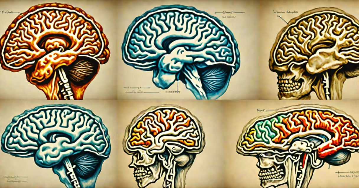
A MRI with contrast uses a injected dye called gadolinium to make certain tissues or abnormalities appear brighter on images, while MRI without contrast relies solely on natural differences in tissue water content and magnetic properties. The contrast agent highlights blood vessels, inflammation, tumors, and infections more than standard scans.
MRI with contrast is often used when detecting small lesions, assessing tumor spread, or evaluating treatment response. MRI without contrast remains the standard for initial screening of joints, brain structure, and spinal cord issues. Both methods use useful magnets and radio waves, but the addition of contrast changes how signals are captured and interpreted.
The choice between the two depends on clinical need. A doctor may order a non-contrast MRI for a routine knee injury or suspected stroke. They'll request contrast if there's concern about cancer, infection, or blood-brain barrier disruption. The decision is based on patient history, symptoms, and imaging goals.
MRI detects hydrogen atoms in water and fat molecules. When exposed to a strong magnetic field and radio pulses, these atoms emit signals that computers convert into detailed cross-sectional images. The strength and timing of these signals depend on tissue type.
In a non-contrast MRI, differences in signal intensity arise from natural variations in tissue composition. For example, fluid-filled cysts appear dark, fatty tissue appears bright, and solid organs show intermediate shades. This allows detection of structural changes like herniated discs or brain bleeds.
When contrast is added, typically through an IV injection, the gadolinium-based agent alters local magnetic fields. It shortens the relaxation time of nearby protons, increasing their signal. Areas with leaky blood vessels-such as tumors or inflamed tissue-absorb more contrast and appear brighter. This enhances visibility of subtle abnormalities invisible on non-contrast scans.
There are two primary MRI scan types based on contrast use. Each serves distinct diagnostic purposes.
| Scan Type | Contrast Agent Used? | Best For | Typical Use Cases |
|---|---|---|---|
| Non-contrast MRI | No | Structural detail, anatomy | Brain tumors, spinal stenosis, joint injuries, stroke evaluation |
| Contrast-enhanced MRI | Yes (gadolinium) | Abnormal tissue activity, blood flow | Cancer staging, infection detection, multiple sclerosis flare-ups |
For example, a non-contrast brain MRI might show a 1.5 cm mass in the frontal lobe. Without contrast, it's hard to tell if it's a benign cyst or a malignant glioma. After contrast injection, the lesion shows intense ring enhancement-indicating disrupted blood-brain barrier typical of aggressive tumors. This difference changes the next steps in care.
Contrast agents are not always needed. A non-contrast MRI of the knee can detect a torn meniscus with 95% accuracy, according to a 2021 study published in Radiology. But for soft-tissue sarcomas, contrast increases sensitivity from 78% to 92%.
Using contrast improves diagnostic confidence. A 2020 review in European Radiology found that contrast-enhanced MRI increased detection rates for metastatic disease in breast cancer patients by 34% compared to non-contrast scans.
However, contrast isn't risk-free. Gadolinium can deposit in the brain and bones, though long-term effects remain uncertain. The FDA warns against using it in patients with severe kidney impairment due to rare but serious conditions like nephrogenic systemic fibrosis. Non-contrast MRI avoids these risks entirely.
Another key distinction: timing. A non-contrast scan takes 20-45 minutes. Adding contrast requires a delay of 5-10 minutes after injection to allow distribution, then additional sequences. Total scan time increases by 10-15 minutes.
Not all institutions offer contrast MRI. Smaller clinics may lack the equipment or trained staff to administer intravenous contrast safely. In such cases, non-contrast MRI becomes the default option-even when contrast would improve accuracy.
Many assume that "MRI with contrast" means a better or more complete scan. That's not true. A non-contrast MRI can provide sufficient detail for many conditions. Overuse of contrast exposes patients to unnecessary risk.
Another myth: contrast makes all tumors visible. In reality, some tumors-especially low-grade gliomas-don't improve even with contrast. Their cells don't disrupt the blood-brain barrier enough to take up the dye. These require specialized sequences like diffusion-weighted imaging, which don't rely on contrast.
Also, contrast doesn't replace other imaging tools. A CT scan with iodine contrast is still preferred for lung nodules or acute trauma. MRI with contrast excels in soft-tissue evaluation, especially in the brain, spine, and abdomen.
I once observed a patient with persistent headaches. Initial non-contrast MRI showed no mass. After contrast, a small dural-based meningioma became visible-smaller than 1 cm, but growing. Without contrast, it would have gone undetected. This case illustrates how context matters: a normal non-contrast scan doesn't rule out pathology.
Doctors base decisions on symptom duration, prior imaging, and suspected diagnosis. For instance:
For children, non-contrast MRI is preferred unless absolutely necessary. The American College of Radiology recommends avoiding gadolinium in pediatric patients under 12 unless clinically justified, due to limited safety data.
Patients should discuss the trade-offs with their radiologist. Risks include allergic-like reactions (1 in 1,000), mild nausea, and rare kidney complications. Benefits include earlier detection, accurate staging, and better treatment planning.
Understanding what is a MRI with and without contrast helps patients make informed choices. Non-contrast MRI provides high-resolution anatomical detail without injections. Contrast-enhanced MRI adds functional insight by revealing abnormal vascular permeability. The two are complementary, not interchangeable.
Always ask your provider why a specific scan type was ordered. If contrast is recommended, understand the reason and potential side effects. Not every scan needs contrast-and sometimes, less is more.