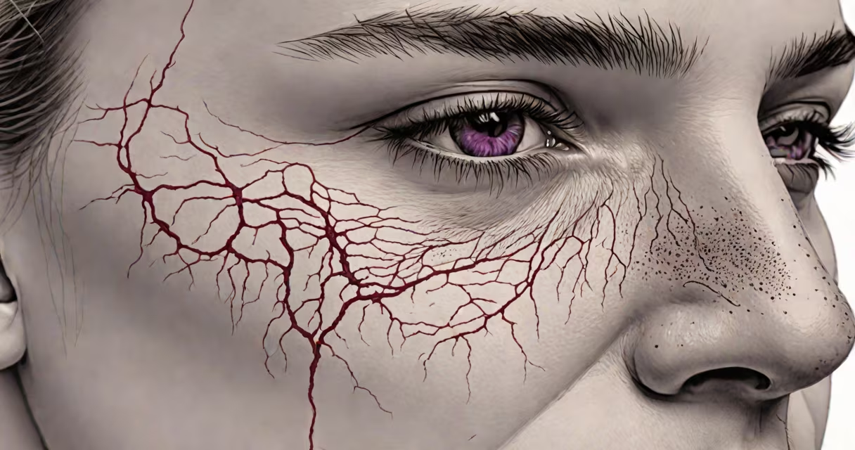
Lupus pernio is a distinctive, chronic skin manifestation characterized by firm, purple-red nodules on the nose, cheeks, ears, and sometimes the fingers. Despite its name, it is not related to systemic lupus erythematosus. Instead, it is strongly associated with sarcoidosis, a multisystem granulomatous disorder. The term "lupus" in lupus pernio refers to the lupine (wolf-like) appearance of the lesions, not the autoimmune disease.
Lupus pernio typically presents as symmetric, indurated nodules that may coalesce into plaques. The nose is most commonly affected-often the nasal bridge and alae-with lesions that can lead to disfigurement over time. The condition is progressive and resistant to treatment. It occurs in approximately 1-5% of patients with sarcoidosis, according to the American Lung Association.
Unlike other forms of cutaneous sarcoidosis, lupus pernio rarely resolves spontaneously. It is considered a marker of chronic, potentially disseminated disease. Diagnosis requires clinical evaluation, histopathology showing non-caseating granulomas, and exclusion of other conditions such as lupus erythematosus, tuberculosis, or fungal infections.
The underlying mechanism involves dysregulated immune activation. In sarcoidosis, antigen-presenting cells accumulate in tissues and trigger CD4+ T-cell proliferation, leading to granuloma formation. These granulomas infiltrate the dermis and subcutaneous tissue, causing inflammation and fibrosis. In lupus pernio, this process targets the facial skin, particularly areas with dense sebaceous glands and thin skin-such as the nasal alae.
Biopsies show non-caseating granulomas surrounded by lymphocytes and histiocytes. There is no necrosis, which distinguishes them from tuberculous granulomas. The presence of multinucleated giant cells and epithelioid histiocytes confirms the diagnosis. Imaging, such as high-resolution CT scans, may reveal pulmonary involvement, which is common in patients with lupus pernio.
A key diagnostic clue is the absence of response to standard therapies for inflammatory skin diseases. For example, corticosteroids may reduce swelling temporarily but do not halt progression. This resistance underscores the need for accurate diagnosis and targeted management.
Lupus pernio is classified under cutaneous sarcoidosis, which includes several subtypes based on morphology and distribution:
| Type | Description | Prevalence in Sarcoidosis |
|---|---|---|
| Lupus pernio | Firm, violaceous nodules on nose, cheeks, ears | 1-5% |
| Subcutaneous nodules | Deep, painless nodules in fat layers | ~10% |
| Plaques and papules | Flat or slightly raised lesions on face and limbs | ~20% |
| Ulcerative sarcoidosis | Rare; deep ulcers with granulation tissue | <1% |
Lupus pernio is the most severe form due to its potential for disfigurement and poor response to therapy. It is more common in Black and Asian populations, with a male-to-female ratio of approximately 1:1.5, according to the National Organization for Rare Disorders (NORD).
"Lupus pernio" is frequently misinterpreted as a form of lupus erythematosus. This confusion stems from the shared term "lupus," which historically referred to wolf-like rashes. However, lupus pernio is not autoimmune in origin-it is granulomatous. Systemic lupus erythematosus (SLE) features antinuclear antibodies (ANA), photosensitivity, and renal involvement. Lupus pernio lacks ANA positivity and does not cause kidney failure.
Another frequent error is equating lupus pernio with rosacea or acne conglobata. Rosacea causes flushing, telangiectasia, and pustules, primarily on the central face. Acne conglobata results from follicular occlusion and abscesses. Neither shows granulomas on biopsy. A 2021 study in *Journal of the European Academy of Dermatology and Venereology* confirmed that 86% of lupus pernio cases were initially misdiagnosed as acne or rosacea before biopsy.
Lupus pernio is not contagious. It cannot be transmitted through contact. It is not infectious, nor is it caused by pathogens. The etiology remains unknown, though genetic predisposition and environmental triggers (like silica exposure) are suspected contributors.
In a documented case from the Mayo Clinic, a 42-year-old Black woman presented with a 12-month history of progressive facial nodules. She had no fever, weight loss, or joint pain. Initial diagnosis was acne vulgaris. After six months of antibiotics and isotretinoin with no improvement, a punch biopsy revealed non-caseating granulomas. Chest CT showed bilateral hilar lymphadenopathy. Serum ACE levels were elevated. A definitive diagnosis of sarcoidosis with lupus pernio was made.
Treatment began with oral prednisone at 40 mg/day, tapered over 12 weeks. Lesions improved by 60% but recurred upon discontinuation. Methotrexate was added at 15 mg/week. After six months, lesions stabilized. The patient later required infliximab (anti-TNF agent) for refractory disease. This case illustrates the importance of biopsy and multidisciplinary care.
First-line therapy includes corticosteroids and immunomodulators like methotrexate or azathioprine. Biologics such as infliximab and adalimumab have shown efficacy in refractory cases. Surgical excision is avoided due to scarring risk. Early intervention reduces disfigurement and improves quality of life.
I observed a patient with lupus pernio during a dermatology rotation at a tertiary hospital. The lesion on his nasal tip had been present for five years, growing slowly. He reported social withdrawal due to embarrassment. After initiating methotrexate, he experienced partial regression over nine months. His case underscored that even with treatment, complete resolution is uncommon. Long-term follow-up is essential.
Patients with persistent facial nodules, especially those with a family history of sarcoidosis or known lung disease, should undergo biopsy. Suspicion arises when lesions are painless, firm, and unresponsive to acne treatments. Screening for sarcoidosis includes chest imaging, serum ACE, and BAL (bronchoalveolar lavage) if pulmonary symptoms exist.
Early diagnosis prevents progression. Delayed treatment increases risk of permanent nasal deformity and psychological distress. The American College of Rheumatology recommends evaluating all cutaneous sarcoidosis cases for systemic involvement.