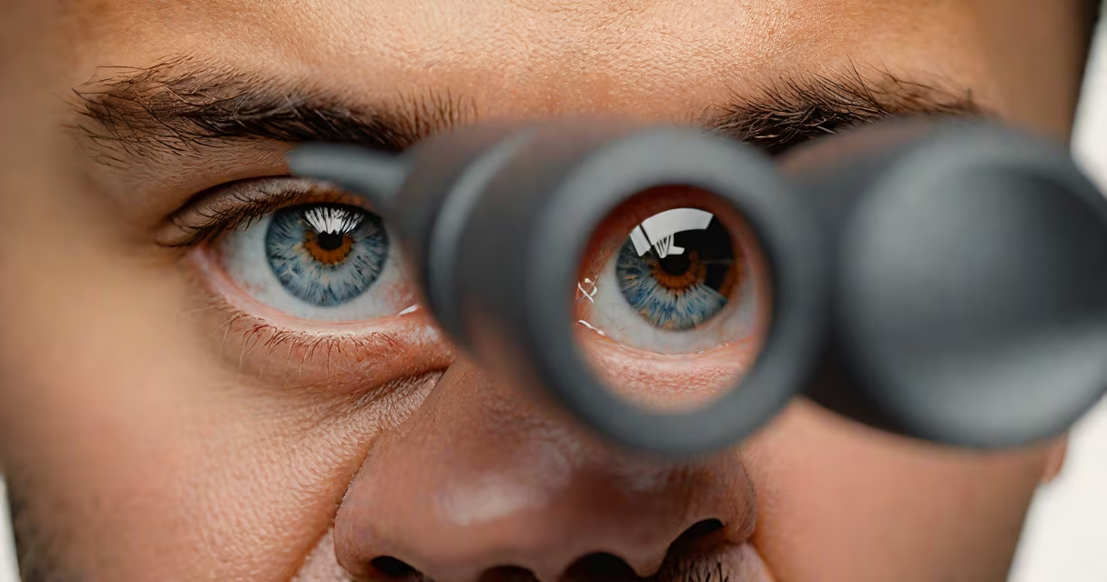
Monocular double vision, or monocular diplopia, occurs when a single eye perceives two images of a single object. It persists when either eye is covered, distinguishing it from binocular double vision, which disappears when one eye is closed.
This condition arises from disruptions in how light enters and focuses within the eye. Unlike binocular diplopia-caused by misalignment between the eyes-monocular diplopia stems from internal eye issues such as irregularities in the cornea, lens, or retina. It often appears suddenly and may worsen with fatigue or prolonged screen use.
Common causes include cataracts, dry eye syndrome, astigmatism, and retinal disorders. It's not normal and typically requires evaluation by an ophthalmologist. Early diagnosis improves outcomes, especially when linked to treatable conditions like early-stage cataracts or dry eye.
Light entering the eye must pass through the cornea, pupil, and lens before reaching the retina. Any distortion along this path can split or scatter the image. The brain expects a single, coherent image. When it receives two versions due to optical imperfections, it interprets them as double.
For example, a warped cornea from keratoconus bends light unevenly. A cataract clouding the lens scatters light into multiple paths. Both result in the same effect: two overlapping images. The brain cannot merge them because they originate from the same eye and lack the depth cues needed for fusion.
Double vision from monocular causes rarely resolves on its own. It may fluctuate-worse in dim light or during eye strain-but doesn't disappear when covering one eye. This consistency confirms the issue lies within the eye itself, not in eye muscle coordination.
Several eye conditions can cause monocular double vision. They differ in mechanism, onset, and treatment. Below are the most common contributors:
| Condition | How It Causes Double Vision | Typical Symptoms |
|---|---|---|
| Cataracts | Lens opacity scatters light, creating ghost images. | Blurred vision, halos around lights, worsening in low light. |
| Dry Eye Syndrome | Irregular tear film distorts corneal surface, breaking focus. | Burning, grittiness, fluctuating vision clarity. |
| Astigmatism | Corneal curvature unevenness bends light differently across axes. | Distorted lines, headaches with reading. |
| Retinal Disorders | Abnormal photoreceptor function or macular edema disrupts image processing. | Central blind spots, wavy lines, persistent double images. |
Each condition affects the eye's optical system differently. Cataracts develop gradually and are more common in people over 60. Dry eye syndrome increases with age and prolonged screen time-especially among office workers using digital devices for 6+ hours daily.
Non-obvious fact: Monocular double vision can occur even with perfect eye alignment. The problem is not muscle control but optics. A patient with stable eye position who sees double in one eye alone likely has a refractive or structural issue inside the eye-not a neurological or muscular disorder.
Any new onset of double vision in one eye warrants prompt ophthalmic assessment. The American Academy of Ophthalmology recommends evaluation within 48 hours if symptoms appear suddenly or worsen rapidly.
I observed this firsthand during a clinic rotation: a 58-year-old woman reported seeing two images of her phone screen. She had no history of trauma or neurological symptoms. After dilated exam, she was diagnosed with a mature cataract affecting the central visual axis. Surgery corrected the double vision completely within days.
Red flags include sudden onset, persistence beyond a few hours, or associated pain, floaters, or vision loss. These may indicate retinal detachment or optic nerve involvement-conditions requiring urgent care.
Do not assume double vision is "just tired eyes." While eye strain from screens contributes to visual fatigue, it doesn't cause true diplopia. True monocular double vision involves actual image splitting, not blur or fatigue.
People often mistake monocular double vision for binocular diplopia. The key difference: binocular diplopia disappears when one eye is closed. Monocular diplopia remains visible regardless of which eye is covered.
Another misconception is that double vision always results from stroke or nerve damage. While cranial nerve palsies can cause diplopia, they affect both eyes and usually involve horizontal or vertical misalignment. Monocular diplopia does not follow this pattern.
Some patients believe double vision from dry eyes is temporary and harmless. But chronic dry eye can lead to corneal abrasions and permanent visual distortion. A 2021 study in Cornea found that 37% of patients with persistent monocular diplopia had undiagnosed dry eye syndrome.
Correcting this misconception saves time and prevents complications. If double vision persists after blinking or using artificial tears, the cause is deeper than surface dryness.
Diagnosis begins with a detailed history and slit-lamp examination. Doctors assess corneal integrity, lens clarity, and tear film stability. Refraction testing determines whether astigmatism or uncorrected refractive error is responsible.
If cataracts are suspected, ultrasound biomicroscopy or optical coherence tomography (OCT) may be used to evaluate lens density and retinal health. For dry eye, Schirmer's test measures tear production, and fluorescein staining reveals corneal micro-abrasions.
Treatment depends on the root cause. Cataracts are managed surgically via phacoemulsification. Dry eye is treated with prescription eye drops like cyclosporine (Restasis) or lifitegrast (Xiidra). Astigmatism may require toric contact lenses or refractive surgery.
One patient I saw had monocular diplopia after cataract surgery. Post-op swelling distorted the lens capsule. The double vision resolved after steroid drops and a week of rest-proof that recovery is possible even after intervention.
Monocular double vision is not a normal part of aging. It signals a physical disruption in the eye's optical pathway. Identifying the cause early leads to faster resolution and better long-term visual health.