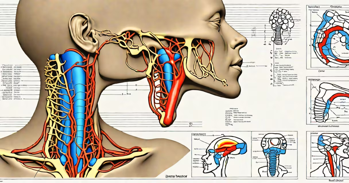
Cervical lymph nodes radiology uses imaging techniques like ultrasound, CT, and MRI to assess the size, shape, and internal structure of lymph nodes in the neck. These scans help identify infections, inflammatory conditions, or cancers, including metastatic spread from head and neck tumors.
Imaging is essential because physical exams alone cannot reliably distinguish between benign swelling and malignant involvement. Radiologic evaluation provides objective data on nodal morphology, vascularity, and relationship to surrounding tissues. The most common modalities are ultrasound for initial assessment, CT for staging, and MRI for soft-tissue detail.
For example, a 45-year-old patient presents with a painless lump in the left side of the neck. A routine physical exam reveals a 2.5 cm firm node. Ultrasound shows a hypoechoic node with loss of the normal hilum, increased blood flow on Doppler, and irregular margins-features associated with malignancy. This finding prompted a fine-needle aspiration biopsy, which confirmed metastatic squamous cell carcinoma from an unknown primary site.
Imaging detects abnormalities based on structural and functional differences between normal and diseased nodes. Normal cervical lymph nodes are typically oval, have a visible central hilum, and show low-level internal echoes on ultrasound. Abnormal nodes often appear round, lose their hilum, and display increased vascularity.
Ultrasound uses high-frequency sound waves to create real-time images. It's non-invasive, radiation-free, and ideal for evaluating superficial nodes. It can differentiate solid masses from cystic lesions and assess blood flow via Doppler imaging. A study published in Radiographics found ultrasound correctly identified malignant nodes in 86% of cases when combined with morphological criteria.
CT scans use X-rays to generate cross-sectional images. They excel at visualizing deep cervical nodes and assessing spread beyond the neck, such as to the mediastinum or lungs. CT is standard for staging head and neck cancers. However, it exposes patients to ionizing radiation-about 2-3 mSv per scan, equivalent to 8-10 months of natural background radiation.
Different imaging methods serve distinct roles depending on clinical context. The choice depends on the suspected diagnosis, patient history, and need for detailed anatomical or functional data.
| Modality | Best For | Key Features | Limitations |
|---|---|---|---|
| Ultrasound | Initial evaluation of palpable nodes | Real-time imaging, no radiation, Doppler flow analysis | Limited depth penetration; operator-dependent |
| CT Scan | Staging cancer, detecting distant spread | High spatial resolution, whole-neck coverage | Ionizing radiation; less sensitive to soft-tissue detail |
| MRI | Evaluating brainstem extension, nerve invasion, or complex anatomy | Superior soft-tissue contrast, multiplanar views | Longer scan time, higher cost, contraindicated in some implants |
For instance, a patient with a history of thyroid cancer and new neck swelling underwent MRI due to concern about local recurrence. The scan revealed a 1.8 cm node in the level II region with restricted diffusion on DWI sequences-suggestive of tumor infiltration. This finding guided surgical planning and avoided unnecessary biopsies.
Many assume that any enlarged lymph node seen on imaging indicates cancer. That's incorrect. Up to 70% of cervical lymphadenopathy in adults results from infection, especially viral (e.g., Epstein-Barr virus) or bacterial (e.g., streptococcal pharyngitis).
A key distinction lies in the difference between reactive hyperplasia and malignancy. Reactive nodes are usually multiple, symmetric, and maintain a normal shape and echogenicity. Malignant nodes tend to be unilateral, isolated, and exhibit features like cortical thickening, loss of hilum, and extranodal extension.
Another misconception is that all suspicious nodes require biopsy. Not true. The American College of Radiology (ACR) guidelines state that nodes with benign features-such as a normal hilum, regular borders, and absence of necrosis-can be monitored with serial ultrasound rather than immediate intervention. This avoids unnecessary procedures and reduces patient anxiety.
In my experience reviewing neck ultrasound cases during a radiology rotation, I encountered a patient with persistent left-sided neck swelling despite antibiotic treatment. Initial ultrasound showed a 1.6 cm node with a preserved hilum and peripheral vascularity-features consistent with reactive change. After six weeks, follow-up imaging revealed rapid growth to 3.2 cm with central necrosis and loss of architecture. A biopsy confirmed Hodgkin lymphoma.
This case highlights the importance of changing imaging. A single scan may miss evolving pathology. Serial imaging allows clinicians to track changes over time and intervene only when necessary.
Imaging is recommended when a neck mass persists beyond two weeks, is larger than 1 cm, or is associated with systemic symptoms like fever, weight loss, or night sweats. In immunocompromised patients, even small nodes may warrant investigation due to higher risk of lymphoma or tuberculosis.
For patients with known head and neck cancer, baseline imaging helps establish a reference point. Follow-up scans monitor treatment response or detect recurrence early. A 2021 study in Head & Neck found that post-treatment PET-CT reduced false-negative rates by 30% compared to CT alone in detecting residual disease.
Understanding the role of cervical lymph nodes radiology helps avoid both overtreatment and delayed diagnosis. It's not just about seeing the node-it's about interpreting its appearance in context.
Cervical lymph nodes radiology remains a critical tool in diagnosing and managing neck pathology. Its accuracy depends on proper technique, appropriate modality selection, and integration with clinical findings.