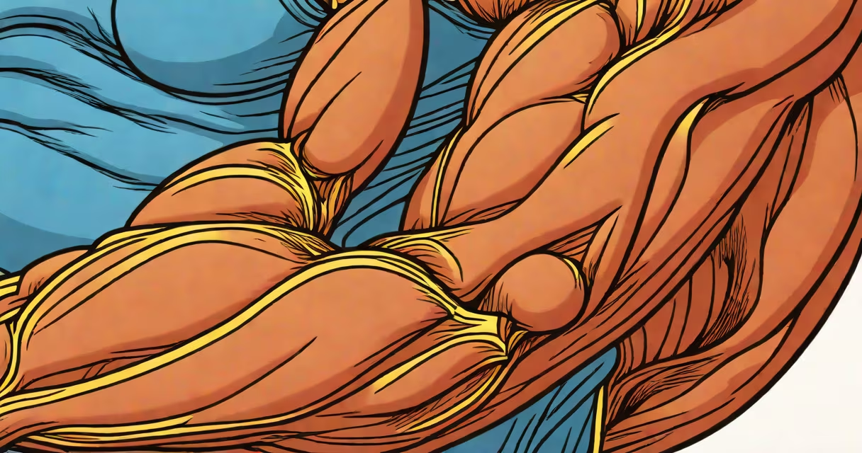
The muscle under skin is the layer of skeletal muscle located just beneath the dermis and subcutaneous fat. It contracts to produce movement, maintain posture, and generate heat. This layer is essential for voluntary motion and is directly connected to bones via tendons.
Skeletal muscles form the bulk of the body's musculature. They lie deep to the skin and fat, separated from the surface by connective tissue layers. When you flex your bicep, you're seeing the contraction of muscle under skin. These muscles are controlled by the somatic nervous system and respond instantly to conscious commands.
Each muscle fiber is surrounded by a thin sheath called the endomysium. Groups of fibers bundle together into fascicles, which are wrapped in perimysium. The entire muscle is enclosed in epimysium, a dense connective tissue layer. This layered structure provides strength and flexibility while allowing precise control during movement.
Muscles under skin contract when motor neurons send electrical signals from the spinal cord. Calcium ions release inside muscle fibers, enabling actin and myosin filaments to slide past each other-a process known as the sliding filament theory. This shortens the muscle, pulling on tendons and moving bones.
For example, when you lift a book, the biceps brachii contracts. The muscle under skin in your upper arm shortens, rotating the forearm upward. This action requires coordination between the brain, spinal cord, and peripheral nerves. Without proper neural input, even strong muscles cannot move effectively.
Every contraction consumes energy. Muscles rely on ATP produced through aerobic and anaerobic metabolism. During intense exercise, lactic acid builds up, causing the burning sensation in muscles under skin. This is a normal byproduct of energy production when oxygen supply lags behind demand.
There are over 600 skeletal muscles in the human body, grouped by location and function. Major groups include:
| Group | Primary Function | Example Locations |
|---|---|---|
| Extensors | Extend limbs | Triceps, quadriceps |
| Flexors | Bend limbs | Biceps, hamstrings |
| Rotators | Rotate joints | Supraspinatus, infraspinatus |
| Stabilizers | Hold posture | Core muscles (transverse abdominis) |
Some muscles under skin are visible through the skin, especially in lean individuals. The rectus abdominis forms the "six-pack" when well-developed. The deltoids create the rounded shoulder contour. Others, like the deep hip rotators, remain hidden beneath layers of fat and fascia.
Not all muscles under skin are visible. The palmaris longus, present in about 14% of people, is often absent. Its absence doesn't affect hand function but makes the wrist appear less defined. This variation shows that muscle presence and visibility aren't always linked.
Muscle under skin can be strained or torn during sudden movements, heavy lifting, or repetitive stress. A strain involves stretching or tearing muscle fibers. A tear occurs when fibers rupture completely. Both cause pain, swelling, and reduced function.
A common mistake is confusing muscle under skin with skin itself. The skin is the outermost barrier. Beneath it lies subcutaneous fat, then the fascia-the connective tissue sheet that encloses muscles. The muscle under skin sits below fascia. Separating these layers is critical for accurate diagnosis and treatment.
Another misconception is that muscle under skin heals quickly. In reality, recovery takes days to weeks. According to the American Academy of Orthopaedic Surgeons, most mild strains resolve in 2-4 weeks with rest and ice. Severe tears may require surgery and months of rehabilitation.
I once observed a runner who ignored persistent calf tightness. After five days of running, he experienced sharp pain and swelling. An MRI confirmed a partial tear in the gastrocnemius muscle-part of the muscle under skin in the lower leg. He stopped activity, applied cold packs, and rested. By week three, mobility improved. This case highlights that ignoring early symptoms leads to prolonged recovery.
People often confuse muscle under skin with tendons, ligaments, or fat. Tendons connect muscle to bone. Ligaments connect bone to bone. Fat lies above the muscle under skin. Fascia surrounds and separates muscles.
For example, the Achilles tendon connects the calf muscles to the heel bone. If you press your finger into the back of your ankle, you feel the tendon. The muscle under skin lies higher up, in the calf region. Confusing them leads to incorrect self-treatment.
Also, muscle under skin differs from visceral muscle. Visceral muscles are involuntary and found in organs like the stomach and intestines. Skeletal muscles under skin are voluntary and responsible for movement. Mixing these categories causes misunderstanding of bodily function.
According to the National Institutes of Health, over 30% of sports injuries involve muscle under skin. Most occur during sudden acceleration or deceleration. Proper warm-up reduces risk by increasing blood flow and elasticity in muscle under skin.
Rest, ice, compression, and elevation (RICE) are standard initial steps for muscle under skin injuries. Apply ice for 15-20 minutes every 2-3 hours during the first 48 hours. Avoid heat and massage during acute phases, as they increase swelling.
After inflammation subsides, gentle stretching and strengthening resume. A study published in the Journal of Athletic Training found that progressive resistance training reduced recurrence rates by 40% in athletes with prior muscle strains.
Prevention includes changing warm-ups before activity and cooldown stretches afterward. Warming up increases muscle temperature, improving contractile efficiency. Cooling down promotes blood flow and removes metabolic waste.
If pain persists beyond a week, seek medical evaluation. Imaging such as ultrasound or MRI can detect tears invisible to the naked eye. Early intervention prevents chronic issues.