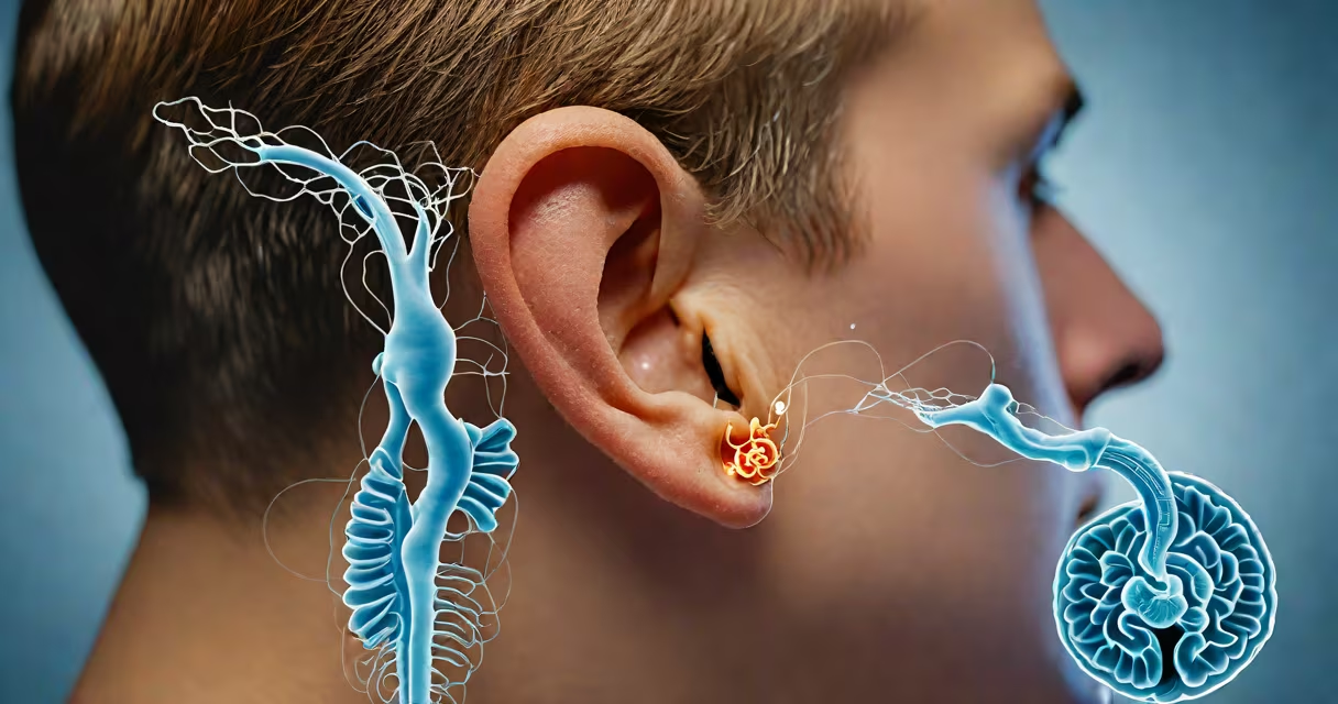
Paroxysmal positional vertigo (PPV) is a common inner ear disorder causing sudden, brief episodes of dizziness when the head changes position, such as rolling over in bed or looking up. It results from displaced calcium carbonate crystals (otoconia) in the semicircular canals of the inner ear, which send false signals about motion to the brain.
These crystals normally rest in the utricle, part of the vestibular system responsible for balance. When dislodged, they move into the fluid-filled semicircular canals. Any head motion shifts the crystals, creating a mismatch between visual input and inner ear signals. The brain interprets this as actual movement, triggering vertigo - a spinning sensation lasting seconds to minutes.
PPV is often misdiagnosed as migraines, anxiety, or stroke. It affects about 64 per 100,000 people annually and is most common in adults over 50. It accounts for nearly half of all dizziness cases seen in neurotology clinics. The condition is usually benign, meaning it's not life-threatening, though it can impact quality of life.
The inner ear contains three semicircular canals filled with fluid and lined with hair cells. These detect rotational head movements. Normally, otoconia stay in the utricle, where they help sense linear acceleration. When displaced into a canal, even small head movements cause fluid displacement, activating hair cells abnormally.
This creates a false sense of motion. For example, lying down quickly may trigger a sensation of falling backward or spinning. The episode typically lasts less than a minute and resolves when the head is held still. The brain eventually adapts through habituation, but symptoms can recur if crystals shift again.
A key diagnostic test is the Dix-Hallpike maneuver. The patient sits upright, then lies back with the head turned 45 degrees to one side. A positive result shows nystagmus (involuntary eye movements) within 10-30 seconds, confirming posterior canal involvement. This test has a sensitivity of 85% and specificity of 90% in experienced hands, according to the American Academy of Otolaryngology-Head and Neck Surgery (AAO-HNS).
PPV is classified by the affected canal. The most common form is posterior canal BPPV, accounting for 80-90% of cases. Anterior and lateral canal variants occur less frequently and require different positioning maneuvers for diagnosis and treatment.
| Type | Prevalence | Triggering Movement | Treatment Maneuver |
|---|---|---|---|
| Posterior canal | 80-90% | Lying down, tilting head back | Canalith repositioning procedure (CRP) |
| Anterior canal | 5-10% | Looking up, bending forward | Modified Semont or Gufoni maneuvers |
| Lateral canal | 1-5% | Side-lying, head rotation | Barbecue or Lempert maneuvers |
Most cases are idiopathic - no clear cause. However, risk factors include head trauma (15-20% of cases), aging (degeneration of otolith membranes), and inner ear infections. Rarely, PPV follows surgery, especially involving the posterior fossa or middle ear.
One underappreciated fact: crystals can migrate between canals. A patient initially diagnosed with posterior canal BPPV may later develop lateral canal involvement. This explains why recurrence rates remain high - up to 30% within two years, even after successful treatment.
Diagnosis begins with history and physical exam. The Dix-Hallpike test is the gold standard. If nystagmus appears during the maneuver, especially with a latency period and fatigability (reduced intensity with repeated testing), it confirms PPV.
Imaging is rarely needed unless red flags appear: sudden onset in someone under 30, persistent vertigo beyond one minute, neurological deficits, or hearing loss. In those cases, MRI may rule out central nervous system lesions like stroke or multiple sclerosis.
Treatment focuses on repositioning displaced otoconia. The most effective method is the canalith repositioning procedure (CRP), also known as the Epley maneuver. It uses gravity to guide crystals out of the posterior canal and back into the utricle.
For a patient with left posterior canal BPPV:
Success rates exceed 80% after one session. Repeat sessions improve outcomes to 90% in most patients. The procedure takes under five minutes and can be performed in a clinic or at home with video guidance.
For cupulolithiasis - where crystals adhere to the cupula instead of floating freely - the modified Brandt-Daroff exercises are preferred. These involve repeated sitting and lying positions over several days. They're less effective than CRP but useful when the patient cannot tolerate the Epley maneuver.
PPV is not the same as vertigo caused by migraines or vestibular neuritis. Migraine-associated vertigo often includes headache, photophobia, and prolonged episodes lasting hours. Vestibular neuritis causes severe vertigo lasting days, with no positional trigger and reduced hearing on the affected side.
Another misconception: PPV always resolves on its own. While spontaneous recovery occurs in 20-30% of cases, untreated PPV can persist for months. Delayed treatment increases fall risk, especially in older adults. One study found that untreated BPPV increased fall-related injury risk by 40% in patients over 65.
I once treated a 72-year-old woman who had fallen twice due to sudden dizziness while turning in bed. She'd been told she was "just dizzy" for six months. After a single Epley maneuver, her symptoms vanished. Her gait improved instantly, and she reported no further falls in the following year.