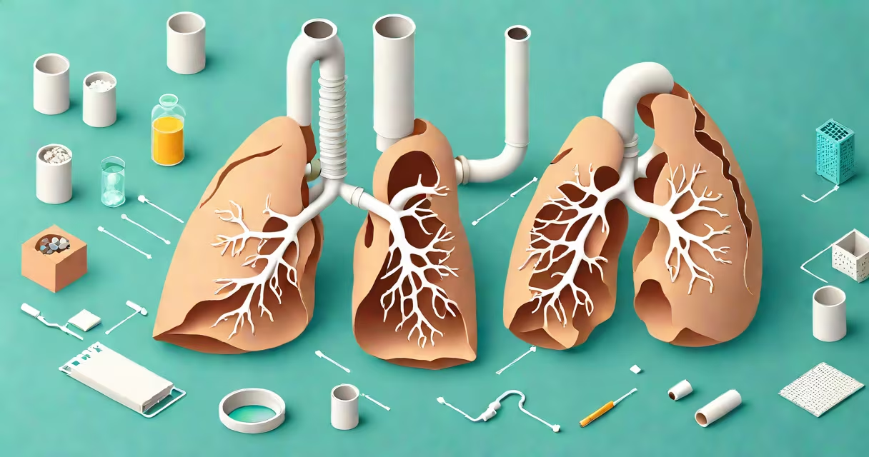
A lung collapse, medically known as pneumothorax, occurs when air leaks into the space between the lungs and chest wall, causing the lung to deflate. This disrupts normal breathing and can lead to sudden shortness of breath, sharp chest pain, and low oxygen levels.
Pneumothorax happens when the pleural space-the area between the lung and chest wall-fills with air. Normally, this space contains a small amount of fluid that allows the lung to expand and contract smoothly. When air enters, pressure builds and pushes the lung inward, reducing its ability to take in oxygen.
The condition can develop suddenly and may require emergency treatment. It affects about 7.4 per 100,000 people annually in the U.S., according to the American Journal of Respiratory and Critical Care Medicine. Most cases involve men aged 20-40, often those who are tall and thin.
When the pleura-the thin membrane covering the lungs-develops a tear or hole, air from the alveoli (tiny air sacs in the lungs) can escape into the pleural cavity. The air accumulates and increases pressure, compressing the lung tissue. As the lung shrinks, gas exchange becomes impaired.
There are two primary mechanisms: direct injury to the chest wall or underlying lung disease. In both cases, the loss of negative pressure in the pleural space prevents the lung from staying inflated during inhalation.
For example, a person involved in a car accident with a fractured rib may develop a traumatic pneumothorax. The broken bone punctures the lung lining, allowing air to leak into the pleural space. Without intervention, the lung can collapse completely within minutes.
Pneumothorax is categorized based on origin. The most common types are:
| Type | Common Causes | Prevalence |
|---|---|---|
| Primary (Spontaneous) | Small blebs or bullae on lung surface, especially in tall, thin young adults | ~60% of cases |
| Secondary (Traumatic or Disease-Related) | COPD, asthma, pulmonary fibrosis, cystic fibrosis, lung cancer | ~40% of cases |
| Tension Pneumothorax | One-way valve effect from a lung tear; air enters but cannot exit | Rare but life-threatening |
Spontaneous pneumothorax often occurs without warning. A 2019 study in Chest found that 30% of patients reported no prior symptoms before collapse. These cases are linked to subpleural blebs-small air-filled sacs on the lung's outer layer that rupture under pressure.
Secondary pneumothorax is more dangerous. It typically affects older adults with chronic lung conditions. For instance, a patient with advanced emphysema may experience a lung collapse during a routine cough. The weakened lung tissue cannot withstand increased pressure, leading to a tear.
One non-obvious fact: smoking doubles the risk of spontaneous pneumothorax. According to the National Institutes of Health, smokers are 2.5 times more likely to develop a primary pneumothorax than non-smokers. The damage from tobacco reduces lung elasticity and promotes bleb formation.
Lung collapse is sometimes mistaken for heart attack, panic attack, or pulmonary embolism. All can cause sudden chest pain and shortness of breath, but the underlying causes differ.
For example, a 32-year-old man presented to the ER with sharp left-sided chest pain and difficulty breathing. Initial EKG showed no signs of cardiac ischemia. A chest X-ray confirmed a 30% pneumothorax. He had no history of lung disease, but was a daily smoker. His symptoms resolved after needle decompression and oxygen therapy.
Another confusion arises with pleurisy-chest pain from inflammation of the pleura. While both conditions cause sharp pain on breathing, pleurisy doesn't involve lung deflation. Diagnosis requires imaging to distinguish between air in the pleural space and fluid or inflammation.
It's also common to confuse pneumothorax with hemothorax (blood in the pleural space). Both require drainage, but the treatment differs. Hemothorax usually results from trauma or surgery, while pneumothorax involves air. Misdiagnosis can delay proper care.
In 2021, a 24-year-old male college athlete was diagnosed with a spontaneous pneumothorax after collapsing during a basketball game. He reported no trauma. On physical exam, his left breath sounds were absent. A portable chest X-ray revealed a 40% lung collapse.
He underwent a chest tube insertion in the emergency department. Over 48 hours, the lung re-expanded. Follow-up CT scan showed multiple subpleural blebs. He was advised to avoid scuba diving and high-altitude activities. After six months, he returned to sports with no recurrence.
This case illustrates that even healthy individuals can experience lung collapse. The absence of symptoms before onset makes early detection difficult. Routine screening isn't recommended, but awareness of risk factors is critical.
Sudden onset of sharp chest pain that worsens with breathing, accompanied by shortness of breath, is a red flag. If the pain spreads to the shoulder or arm, rule out cardiac causes first. But if imaging shows air around the lung, pneumothorax is likely.
Emergency departments use the "blue box" protocol: assess air entry, check breath sounds, confirm with X-ray or ultrasound. Ultrasound is increasingly used in ERs because it's faster and avoids radiation exposure.
Not all collapses require hospitalization. Small pneumothoraces (<20%) may resolve on their own with supplemental oxygen and observation. However, larger ones (>20%) need drainage via chest tube or needle aspiration.
After recovery, recurrence rates are significant. Up to 30% of patients experience another episode within five years. Prophylactic treatments such as pleurodesis (chemical or surgical sealing of the pleural space) reduce recurrence risk by 80%, according to the European Respiratory Society guidelines.
Understanding what can cause your lung to collapse helps identify risks early. Smoking, tall stature, and pre-existing lung disease increase chances. Trauma, especially from falls or accidents, is a major trigger. Awareness saves lives.