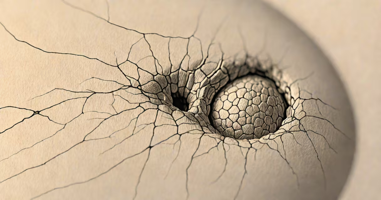
Epidermal cysts develop when keratin-producing cells move deeper into the skin and multiply, forming a sac filled with keratin, a protein found in skin, hair, and nails. These cysts typically appear as slow-growing, round lumps beneath the skin, often with a small dark spot at the center.
They form when the outer layer of skin (epidermis) grows abnormally into deeper tissue. This usually happens after minor trauma, such as a scratch or acne lesion, which disrupts the hair follicle. The displaced cells then create a closed pocket that fills with keratin. Over time, this trapped material hardens into a waxy plug, creating a visible bump.
Most epidermal cysts are benign and do not require treatment unless they become inflamed, infected, or painful. They commonly occur on the face, neck, back, and scalp. While they're not cancerous, they can resemble other skin conditions like lipomas or abscesses, making accurate diagnosis important.
The process begins when the skin's outermost layer, the epidermis, regenerates abnormally. Normally, skin cells grow at the base of hair follicles and gradually move upward, shedding at the surface. When this cycle is disrupted-often by injury or inflammation-the cells may migrate inward instead of outward.
These misplaced cells continue dividing and produce keratin, which accumulates inside a sealed-off sac. As the sac expands, it pushes against surrounding tissue, forming a lump under the skin. The central pore, often visible as a tiny black dot, allows some keratin to escape, giving the cyst a characteristic appearance.
A 2021 study published in the Journal of the American Academy of Dermatology found that 75% of epidermal cysts were associated with prior skin trauma, including acne scarring or minor cuts. This confirms that physical disruption of the follicle is a primary trigger, not just genetic predisposition.
There are two main types: true epidermal cysts and pseudocysts. True epidermal cysts originate from the epidermis and contain keratin. Pseudocysts are not true cysts-they result from fluid accumulation in tissues, often due to infection or inflammation.
Common triggers include:
While most cases are isolated, people with a family history of multiple cysts may have a higher risk. A 2019 review in Dermatologic Clinics noted that individuals with steatocystoma multiplex develop numerous cysts during adolescence, often on the chest and back.
Epidermal cysts are frequently mistaken for sebaceous cysts-a term still used in lay language but technically incorrect. Sebaceous glands produce oil (sebum), and their blockage leads to different lesions. True sebaceous cysts are rare; most so-called "sebaceous" cysts are actually epidermal cysts.
Another confusion arises with abscesses. Both can appear red, swollen, and tender. But abscesses are caused by bacterial infections and fill with pus, while epidermal cysts contain keratin and are sterile unless ruptured. Rupture can cause severe inflammation, mimicking infection.
Some people mistake epidermal cysts for skin cancers like basal cell carcinoma. However, cancerous growths tend to grow rapidly, bleed easily, and lack a central pore. A biopsy is required to confirm malignancy, though this is extremely rare in epidermal cysts.
In 2022, a 34-year-old woman presented with a 1.2 cm cyst on her left upper back. She reported it had been present for five years, slowly increasing in size. The lesion had a central punctum and was firm to touch. Biopsy confirmed it was a true epidermal cyst lined with stratified squamous epithelium and filled with laminated keratin.
She had no history of acne or trauma to the area. However, she recalled a minor burn from a hot iron at age 16. This aligns with clinical evidence: even decades later, prior skin damage can lead to delayed cyst formation.
After surgical excision, the cyst did not recur. The procedure took 20 minutes under local anesthesia. Pathology results showed no signs of malignancy or infection.
Use this comparison to clarify differences:
| Type | Origin | Content | Common Location | Distinctive Feature |
|---|---|---|---|---|
| Epidermal cyst | Epidermis, displaced cells | Keratin, waxy material | Face, neck, back, scalp | Central pore, slow growth |
| Sebaceous cyst | Sebaceous gland | Sebum (oil) | Face, ears, eyelids | Rare; often misnamed |
| Epidermoid cyst | Epidermis, same as epidermal | Keratin | Same as above | Alternate name for same condition |
| Lipoma | Fatty tissue | Adipose cells | Subcutaneous layers | Soft, movable, not connected to follicle |
Epidermal cysts are not related to cholesterol deposits or metabolic disorders. Unlike xanthomas-fatty nodules linked to high cholesterol-epidermal cysts have no connection to blood lipid levels.
I once observed a dermatologist remove a 2.5 cm epidermal cyst from a patient's neck during a clinic visit. The cyst had been present for seven years and recently became tender. After numbing the area, the doctor made a small incision, drained the keratin plug, and removed the entire sac. The procedure took less than ten minutes. The patient reported immediate relief and no recurrence after six months.
This case illustrates that complete removal prevents recurrence. Partial drainage only offers temporary relief, as the sac remains and will refill.
See a healthcare provider if a cyst:
These signs suggest infection or rupture. Untreated, a ruptured cyst can lead to scarring or abscess formation. Surgical removal is the most effective long-term solution.
Understanding what causes epidermal cyst is essential for proper care. Most are harmless and don't need treatment. But knowing the signs of complications ensures timely intervention. Recognizing the role of trauma, genetics, and cellular migration clarifies why these cysts form-and how to manage them effectively.