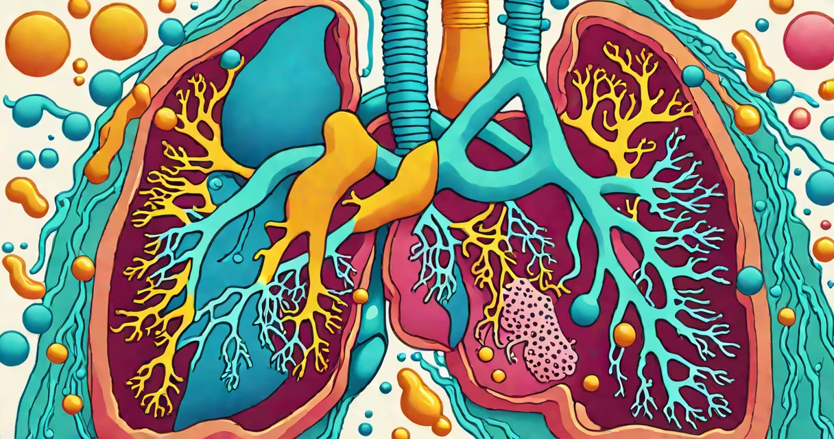
Fluid in your lungs, medically known as pulmonary edema or pleural effusion, happens when excess fluid builds up in the lung tissues or the space between the lungs and chest wall. This disrupts oxygen exchange and can cause shortness of breath, coughing, and fatigue. The underlying cause varies widely and may involve heart, lung, or systemic conditions.
Pulmonary edema results from fluid leaking into the air sacs (alveoli) due to increased pressure in the pulmonary capillaries. This most commonly stems from left-sided heart failure, where the heart cannot pump blood efficiently, causing fluid to back up into the lungs. According to the American Heart Association, acute pulmonary edema affects approximately 1 million people annually in the U.S., with heart failure being the primary driver in over 80% of cases.
Pleural effusion refers to fluid accumulating in the pleural space-the area between the two layers of the pleura that line the lungs and chest cavity. This type is often caused by infections like pneumonia, cancer, or inflammatory diseases such as lupus or rheumatoid arthritis. A 2021 study published in Chest found that malignancy accounted for 15-20% of pleural effusions in adults, while heart failure was responsible for about 30%.
Fluid enters the lungs through two distinct pathways. In pulmonary edema, elevated pressure in the pulmonary veins forces fluid out of capillaries and into alveolar spaces. This process is driven by cardiac dysfunction-especially reduced left ventricular ejection fraction-or conditions like high-altitude pulmonary edema, which occurs in unacclimated individuals ascending rapidly above 2,500 meters.
In pleural effusion, fluid leaks from blood vessels into the pleural cavity due to inflammation, low protein levels in the blood, or blockages in lymphatic drainage. The pleura normally produces a small amount of lubricating fluid, but disease processes can overwhelm this balance. For example, tuberculosis can trigger exudative effusions with high protein content, while nephrotic syndrome leads to transudative effusions with low protein.
A key distinction: pulmonary edema affects lung tissue and impairs gas exchange, while pleural effusion compresses the lung from the outside. Both reduce lung volume, but they require different diagnostic approaches and treatments.
The following table outlines the main categories of fluid in the lungs, their mechanisms, and typical causes:
| Type | Location | Fluid Type | Common Causes |
|---|---|---|---|
| Pulmonary edema | Alveoli and interstitial spaces | Transudate (low protein) | Heart failure, high-altitude exposure, acute kidney injury |
| Pleural effusion | Pleural space | Exudate (high protein) or transudate | Pneumonia, cancer, autoimmune disease, cirrhosis |
Exudative effusions are typically inflammatory or infectious. An example is a patient with community-acquired pneumonia developing a right-sided effusion confirmed via ultrasound and thoracentesis. Analysis of the fluid revealed a high white blood cell count and lactate dehydrogenase (LDH) level-both indicators of exudate.
Transudative effusions result from imbalances in hydrostatic or oncotic pressure. A classic case is a 68-year-old man with congestive heart failure whose chest X-ray shows bilateral blunting of the costophrenic angles-a sign of fluid in the pleural space. His serum albumin is normal, but his central venous pressure is elevated, confirming fluid overload from cardiac strain.
Many assume fluid in the lungs always signals heart failure. While heart-related causes are frequent, they're not universal. For instance, patients with severe sepsis can develop pulmonary edema due to capillary leak syndrome, even without cardiac issues. A 2020 review in Critical Care Medicine noted that sepsis-induced pulmonary edema accounts for nearly 10% of non-cardiogenic cases.
Another confusion arises between pleural effusion and pleurisy. Pleurisy is inflammation of the pleura, often causing sharp pain with breathing. It may lead to effusion, but the two are not synonymous. Pleurisy without fluid is common in viral infections, whereas effusion without pain can occur in silent malignancies.
Not all fluid in the lungs is visible on standard chest X-rays. Small effusions may only show up on ultrasound or CT scans. During a clinical rotation, I observed a patient with advanced ovarian cancer who had a large left-sided effusion on CT but minimal findings on a routine chest X-ray-underscoring the need for imaging beyond basic radiography.
Symptoms like sudden shortness of breath, orthopnea (difficulty breathing lying flat), or paroxysmal nocturnal dyspnea (waking up gasping for air) suggest pulmonary edema and warrant emergency care. These signs indicate acute volume overload and risk of respiratory failure.
For pleural effusion, symptoms include dull chest pain worsened by deep breaths, persistent cough, and reduced exercise tolerance. A patient with a history of breast cancer developed progressive dyspnea and was diagnosed with malignant effusion after a thoracentesis revealed adenocarcinoma cells in the fluid.
Fluid in your lungs is not a single condition but a symptom of multiple pathologies. Understanding the source-cardiac, infectious, neoplastic, or inflammatory-is essential for treatment. Always consult a healthcare provider for accurate assessment and management.