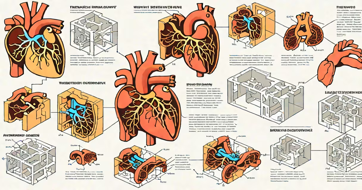
Tetralogy of Fallot is a congenital heart defect caused by four specific structural abnormalities in the heart that develop during early fetal development. These include a large ventricular septal defect, pulmonary stenosis, overriding aorta, and right ventricular hypertrophy. The condition results in oxygen-poor blood mixing with oxygen-rich blood, leading to cyanosis.
These defects form between the 5th and 8th weeks of gestation. The root cause lies in abnormal development of the cardiac neural crest cells and the outflow tract septum. When these structures fail to migrate properly or fuse correctly, the resulting malformations disrupt normal blood flow through the heart.
Genetic factors contribute in about 10-15% of cases. Mutations in genes such as *TBX1* and *NOTCH1* have been linked to the condition. Chromosomal abnormalities like 22q11.2 deletion syndrome (DiGeorge syndrome) occur in approximately 25% of affected individuals. The remaining cases are sporadic with no clear genetic trigger.
The four defining features of tetralogy of Fallot arise from improper alignment of the outflow tract septum. This misalignment prevents the aortic valve from being positioned directly above the left ventricle. Instead, it straddles the ventricular septal defect, allowing blood from both ventricles to enter the aorta.
Pulmonary stenosis restricts blood flow from the right ventricle to the lungs. As a result, deoxygenated blood is shunted from the right ventricle into the left side of the heart via the ventricular septal defect. This mixture reduces arterial oxygen saturation, causing cyanosis-the sign sign of the condition.
Right ventricular hypertrophy develops as a compensatory response to increased pressure in the right ventricle. Over time, the muscle thickens to maintain output despite the obstruction. This adaptation can worsen symptoms if untreated.
Tetralogy of Fallot exists in several forms based on severity and associated anomalies. The most common variant is isolated tetralogy, where all four features are present without additional major cardiac defects.
A rarer form is "pulmonary atresia with tetralogy," where the pulmonary valve is completely closed. This increases the burden on the right ventricle and often requires surgical intervention soon after birth.
| Type | Key Features | Prevalence |
|---|---|---|
| Classic Tetralogy | Large VSD, pulmonary stenosis, overriding aorta, RVH | ~75% |
| Pulmonary Atresia Variant | No pulmonary valve opening; severe obstruction | ~15% |
| With 22q11.2 Deletion | Associated with DiGeorge syndrome; facial dysmorphology | ~25% |
One non-obvious fact: Not all patients with tetralogy of Fallot present with cyanosis at birth. Some remain acyanotic until later infancy due to variable degrees of pulmonary stenosis. A child may appear healthy initially but develop "tet spells" - sudden episodes of severe cyanosis triggered by crying or feeding.
Tetralogy of Fallot is often mistaken for other cyanotic heart diseases such as transposition of the great arteries or truncus arteriosus. However, the presence of a large ventricular septal defect with right-to-left shunting and pulmonary stenosis distinguishes it.
Another misconception is that tetralogy of Fallot is always fatal without surgery. While untreated cases lead to poor outcomes, survival into adulthood is possible with timely repair. The 10-year survival rate after corrective surgery exceeds 95%, according to the American Heart Association.
Some assume it's purely genetic. But the majority of cases are sporadic. Only about 10-15% have a family history. The risk to siblings is less than 1%, unless a chromosomal abnormality is confirmed.
In 2018, a newborn in Boston presented with central cyanosis and a loud systolic murmur. Echocardiography revealed a 1.2 cm ventricular septal defect, marked pulmonary stenosis with a peak gradient of 68 mmHg, and an overriding aorta covering 70% of the ventricular septum. Right ventricular hypertrophy was evident on MRI.
Genetic testing identified a de novo mutation in *TBX1*. The infant underwent complete repair at 3 months old using a transannular patch to close the VSD and relieve pulmonary stenosis. Postoperative recovery was uneventful. By age 2, the child had normal exercise tolerance and no residual shunting.
This case illustrates that while genetics play a role in some cases, surgical correction remains effective even in complex presentations.
I observed a pediatric cardiology team perform a preoperative echocardiogram on a 6-month-old with tet spells. The video loop showed turbulent flow across the pulmonary valve and a jet of blood crossing the VSD into the aorta. The visual confirmation of the anatomical defect made the diagnosis undeniable. That moment underscored how precise imaging is in diagnosing structural heart disease.
Infants with tetralogy of Fallot typically show signs within the first few weeks of life. Cyanosis, especially during feeding or crying, is a red flag. Clubbing of fingers may appear later. Parents should seek evaluation if their baby appears blue, breathes rapidly, or fails to gain weight.
Early diagnosis improves outcomes. The American Academy of Pediatrics recommends screening all newborns with pulse oximetry within 24 hours of birth. A reading below 95% in either hand or foot warrants further investigation.
Tetralogy of Fallot shares etiological pathways with other congenital heart defects involving the outflow tract. The same developmental errors that cause tetralogy can lead to truncus arteriosus or double outlet right ventricle.
Individuals with 22q11.2 deletion syndrome have a 20-30% chance of developing tetralogy of Fallot. This syndrome also includes immune deficiency, hypocalcemia, and characteristic facial features. Early detection allows for coordinated care across multiple specialties.
For those diagnosed with tetralogy of Fallot, follow-up is essential. Long-term complications include arrhythmias, residual pulmonary regurgitation, and late-onset right heart failure. Regular monitoring with echocardiograms and ECGs is recommended throughout life.
Understanding the underlying causes helps guide treatment. Corrective surgery is usually performed between 3 and 6 months of age. The procedure involves closing the VSD and relieving pulmonary stenosis-often with a patch to widen the right ventricular outflow tract.
After successful repair, most patients live normal lives. Physical activity restrictions are minimal. The long-term prognosis is excellent, especially with regular follow-up.
While the exact cause of tetralogy of Fallot remains multifactorial, advances in prenatal imaging and genetic testing allow earlier detection and better outcomes. Awareness of risk factors-such as maternal diabetes, rubella infection during pregnancy, or exposure to certain medications-can inform preventive strategies.
Knowing what causes tetralogy of Fallot enables early diagnosis, informed decision-making, and improved quality of life. With timely intervention, children born with this condition can expect decades of healthy, active living.