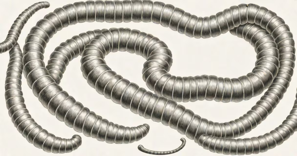
Hookworms in stool appear as thin, coiled, thread-like worms, typically 5-13 millimeters long, with a distinct curved mouthpart resembling a hook. They are visible to the naked eye in fresh fecal samples and indicate active intestinal infection.
Hookworms are parasitic nematodes that attach to the small intestine lining using their oral hooks. They feed on blood, leading to iron deficiency anemia and gastrointestinal distress. The adult worms pass eggs in the stool, which hatch into larvae in warm, moist soil. Infection occurs when larvae penetrate bare skin, usually through walking on contaminated ground.
Diagnosis relies on microscopic detection of hookworm eggs in stool samples. Eggs are oval, about 50-60 micrometers wide, and contain a developing embryo. However, mature adult worms may occasionally be seen in stool during passage, especially in heavy infections. Their presence confirms ongoing infestation and requires treatment.
Adult hookworms resemble pale, translucent threads. When alive, they move slowly and coil tightly. Dead specimens may appear darker or frayed. Their most distinctive feature is the hooked anterior end, visible under a 10x-40x microscope lens. This structure differentiates them from other intestinal worms like roundworms or tapeworms.
Hookworms are not uniformly distributed in stool. A single sample may miss eggs or worms due to intermittent shedding. For accurate diagnosis, multiple stool samples collected over three days are recommended. The Kato-Katz technique, used in tropical medicine, measures egg density per gram of stool to assess infection intensity.
Hookworms are not always visible in stool. Many cases are detected only through microscopy. In rare instances, adults pass through the rectum intact. A 2018 study in the Journal of Clinical Parasitology documented a case where a patient passed a live female *Ancylostoma duodenale* worm in stool after treatment failure, confirming resistance to albendazole.
Hookworms are often mistaken for mucus strands, plant fibers, or hair. Their thin, flexible form resembles intestinal mucus or debris. In low-light conditions or poor sample preparation, they may be overlooked entirely.
Other intestinal parasites differ in appearance:
Hookworms are frequently confused with pinworms due to similar size and shape. However, pinworms lack hooks and are typically found in the lower intestines. Hookworms require a higher degree of clinical suspicion due to their blood-feeding behavior and potential for severe anemia.
A 34-year-old woman in rural Kenya presented with fatigue, pallor, and abdominal cramping. She reported walking barefoot daily on farmland. A stool sample revealed 120 hookworm eggs per gram of feces using the Kato-Katz method. On visual inspection, two coiled, 8-mm-long worms were identified in the sample.
The patient was treated with albendazole (400 mg single dose). Follow-up stool exams showed no eggs or worms after 14 days. Her hemoglobin improved from 8.2 g/dL to 11.8 g/dL within six weeks. This case illustrates that while eggs are the gold standard for diagnosis, adult worms can be visually confirmed in stool during active passage.
According to the World Health Organization (WHO), hookworm affects over 576 million people globally, primarily in tropical and subtropical regions. Transmission is preventable through footwear use and improved sanitation. Routine screening in endemic areas reduces morbidity and prevents chronic anemia.
If you observe thin, coiled, moving worms in your stool-especially with symptoms like fatigue, weight loss, or unexplained anemia-seek medical evaluation. Do not attempt self-diagnosis based on internet images. Worms may resemble debris or tissue fragments.
Stool samples should be submitted fresh, ideally within two hours of collection. Delayed samples degrade parasite morphology. Avoid alcohol-based preservatives unless instructed. Always follow lab guidelines for transport and storage.
For reference, pancreatic stool appears greasy, pale, and foul-smelling due to fat malabsorption. SIBO-related stool is often loose, frothy, and contains mucus. These conditions do not involve parasitic worms.
During a field visit to a clinic in Uganda, I observed a child's stool sample containing three live hookworms. The child had persistent diarrhea and low hemoglobin. The worms were photographed and sent to a central lab for species identification. This confirmed *Necator americanus*, the most common hookworm in the Americas and Africa.
Hookworms in poop are not just a visual curiosity-they signal a treatable parasitic infection. Early detection prevents long-term complications like stunted growth in children and reduced work capacity in adults. If you suspect infection, consult a healthcare provider for proper testing and treatment.
Understanding what hookworm looks like in stool enables faster diagnosis and intervention. Recognizing the thin, coiled, hook-tipped worms helps distinguish them from other intestinal contents. Prompt action reduces transmission and improves outcomes.