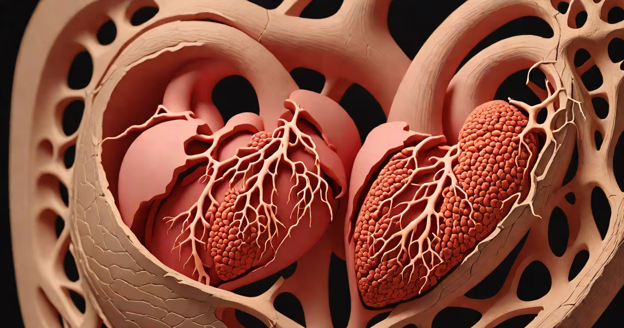
An infarct is a region of dead tissue resulting from interrupted blood supply, typically due to a clot or blockage in an artery. This process, called ischemia, leads to cell death when oxygen and nutrients cannot reach the affected area. The term applies to organs such as the heart, brain, kidneys, and limbs.
Infarcts occur when blood flow stops abruptly. Without oxygen, cells begin to die within minutes. In the heart, this is known as a myocardial infarction-commonly called a heart attack. In the brain, it's a stroke. The size and location determine severity and long-term impact. A small infarct may cause minimal symptoms; a large one can lead to permanent disability or death.
Medical imaging such as MRI or CT scans can detect infarcts by showing areas of abnormal density or lack of perfusion. Blood tests measuring enzymes like troponin rise after a heart infarct, helping confirm the diagnosis. Treatment focuses on restoring blood flow quickly-often through clot-busting drugs or mechanical intervention like angioplasty.
Blood flow interruption begins with atherosclerosis, where fatty deposits build up in arteries. Over time, these plaques can rupture, triggering a blood clot. If the clot blocks a critical vessel, downstream tissue becomes ischemic. After about 20-30 minutes without oxygen, irreversible cell death occurs.
The timeline varies by organ. Brain tissue dies faster than heart muscle-neurons begin dying within 5 minutes of oxygen deprivation. In contrast, skeletal muscle can survive up to 6 hours under low flow conditions. This difference affects emergency response strategies.
A 2021 study published in The Lancet found that early reperfusion therapy reduces mortality in acute myocardial infarction by 35%. Patients treated within 90 minutes of symptom onset had better outcomes than those delayed beyond 3 hours.
Infarcts are categorized by location and underlying mechanism. The most common types include:
| Type | Common Location | Primary Cause | Key Feature |
|---|---|---|---|
| Myocardial | Heart muscle | Coronary artery occlusion | Causes chest pain, ECG changes, elevated troponin |
| Cerebral | Brain | Carotid or cerebral artery blockage | Can cause paralysis, speech loss, vision deficits |
| Pulmonary | Lungs | Clot in pulmonary artery | Often sudden onset dyspnea, pleuritic chest pain |
| Renal | Kidneys | Renal artery thrombosis | May present with flank pain, decreased urine output |
Each type requires rapid identification and targeted treatment. For example, a cerebral infarct must be differentiated from hemorrhagic stroke using a CT scan. Misdiagnosis can worsen outcomes-administering anticoagulants in a bleeding stroke causes fatal complications.
Infarct is often mistaken for atrophy, which involves tissue shrinkage due to disuse or aging-not lack of blood flow. Atrophy results in gradual loss of function, while infarction causes sudden, irreversible damage. The two processes are unrelated despite both involving tissue decline.
Another frequent mix-up is between infarct and necrosis. Necrosis is a broad term for cell death from any cause-trauma, infection, toxins. An infarct is a specific form of necrosis caused by vascular occlusion. Not all necrosis is ischemic; not all ischemia leads to infarction.
I once observed a patient with a leg ulcer mislabeled as "infarct" during a ward round. The lesion was actually due to chronic venous insufficiency. Correct diagnosis required Doppler ultrasound to rule out arterial blockage. This case highlights how precise terminology prevents incorrect treatment paths.
Consider a 62-year-old man presenting with crushing chest pain radiating to his left arm. His ECG shows ST-segment elevation in leads II, III, and aVF. Troponin levels rise above the normal limit. A coronary angiogram reveals a complete blockage in the right coronary artery.
Within 45 minutes, a stent is placed to restore blood flow. The area of dead heart muscle-the infarct-is confirmed via late gadolinium enhancement MRI. The size of the infarct correlates with future risk of heart failure. Follow-up care includes antiplatelets, statins, and cardiac rehab.
This scenario illustrates how infarct detection relies on clinical signs, lab values, and imaging. Early recognition saves lives. Delayed treatment increases infarct size and reduces recovery potential.
Using "infarct" correctly ensures accurate communication among healthcare providers. Mislabeling tissue damage leads to errors in diagnosis, treatment, and research. For example, classifying a tumor-related necrosis as an infarct distorts data on stroke incidence.
According to the World Health Organization, standardized terminology improves global health reporting. The International Classification of Diseases (ICD-11) codes infarcts under chapter 12, "Diseases of the circulatory system," with specific codes for myocardial and cerebral events.
Understanding infarct also aids patients in grasping their condition. Knowing that a "heart attack" means tissue death from blocked blood flow helps them comply with medication and lifestyle changes.