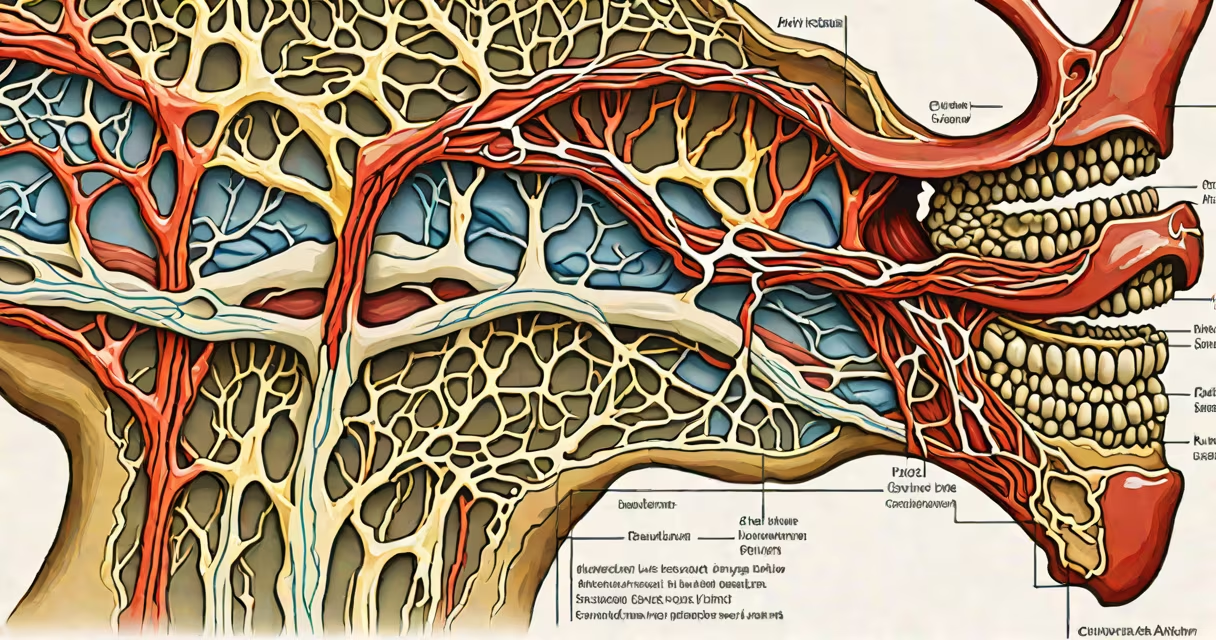
The external carotid artery branches are a network of vessels that deliver oxygenated blood to the face, scalp, neck, and oral cavity. These branches arise from the external carotid artery, which originates from the common carotid artery at the level of the thyroid cartilage.
These arteries form a critical part of the head's circulatory system. They supply tissues involved in mastication, facial expression, and sensory functions. Understanding them is essential for diagnosing and treating conditions such as facial trauma, hemorrhage, and vascular malformations.
Each branch has a distinct anatomical course and function. The most prominent include the superior thyroid, lingual, facial, occipital, posterior auricular, and maxillary arteries. The maxillary artery further divides into terminal branches, including the superficial temporal and middle meningeal arteries.
Blood flows from the common carotid artery into the external carotid artery at the bifurcation of the internal and external carotid arteries. From there, the external carotid artery ascends along the side of the neck, giving off its branches in a consistent sequence.
Each branch follows a predictable path. For example, the facial artery curves around the mandible and supplies the lower lip and nose. The superficial temporal artery runs upward behind the ear and divides into frontal and parietal branches, supplying the scalp.
These vessels operate under pressure gradients maintained by cardiac output and arterial resistance. When blood flow is disrupted-due to trauma, stenosis, or embolism-tissues supplied by these arteries may suffer ischemia or necrosis.
The primary branches of the external carotid artery serve specific regions and have distinct clinical implications.
| Branch | Primary Supply | Clinical Relevance |
|---|---|---|
| Superior thyroid artery | Thyroid gland, larynx, and anterior neck muscles | Used in surgical ligation during thyroidectomy; damage causes hoarseness due to recurrent laryngeal nerve proximity. |
| Lingual artery | Tongue and floor of mouth | Source of bleeding in tongue lacerations; involved in angiographic embolization for postpartum hemorrhage. |
| Facial artery | Face, lips, nasal mucosa | Common site of trauma-related hemorrhage; palpable at the mandibular angle. |
| Occipital artery | Posterior scalp, neck muscles | Supplies the scalp region behind the ear; relevant in migraine surgery targeting greater occipital nerve. |
| Posterior auricular artery | External ear, temporal muscle | Can be injured during ear surgery; contributes to blood supply of the pinna. |
| Maxillary artery | Deep face, sinuses, teeth, brain dura | Terminal branch of external carotid; enters skull via foramen ovale. Supplies the mandible and maxilla. |
| Superficial temporal artery | Temporal scalp, forehead | Site of temporal arteritis; inflammation leads to vision loss if untreated. |
The maxillary artery is not a direct branch but rather a terminal division of the external carotid. It enters the infratemporal fossa and gives rise to multiple intracranial and extracranial branches.
A key distinction often missed: the external carotid artery does not supply the brain directly. That role belongs to the internal carotid artery. The external carotid supports peripheral structures, while the internal carotid handles cerebral perfusion.
These branches are critical in surgical planning. During facial reconstruction or neck dissection, surgeons identify and preserve these vessels to avoid ischemia in important tissues.
For example, in a patient undergoing parotidectomy, the superficial temporal artery lies close to the facial nerve. Damage to this vessel can cause flap necrosis in reconstructive procedures.
Another application is in interventional radiology. Embolization of the facial artery is performed to control epistaxis (nosebleeds) when conservative measures fail. A 2021 study in the Journal of Neurointerventional Surgery reported a 94% success rate in stopping refractory epistaxis using selective catheterization of the facial artery.
The superficial temporal artery is a frequent site for biopsy in suspected giant cell arteritis. A positive result confirms diagnosis and prompts immediate steroid therapy to prevent irreversible vision loss.
Many confuse the external carotid artery branches with those of the internal carotid. The internal carotid supplies the brain via the anterior and middle cerebral arteries. The external carotid supplies the face and scalp.
Another misconception is that all branches are visible on standard imaging. Ultrasound can visualize the facial and superficial temporal arteries, but deeper branches like the maxillary artery require CT angiography or MR angiography.
A non-obvious fact: the posterior auricular artery can sometimes be a collateral pathway in cases of internal carotid occlusion. This rare compensatory mechanism underscores the importance of assessing both systems during stroke evaluation.
I once observed a patient with a facial laceration from a fall. The wound bled profusely despite compression. Doppler ultrasound revealed active flow in the facial artery. After identifying the source, we successfully controlled bleeding with direct suture and intraoperative vasopressin injection.
The external carotid artery branches are essential for perfusing the face, scalp, and neck. Their precise anatomy enables targeted interventions in trauma, surgery, and vascular disease. Recognizing their individual roles prevents complications during medical procedures.
Understanding the external carotid artery branches improves diagnostic accuracy and treatment outcomes. For detailed imaging guidance, refer to this guide.