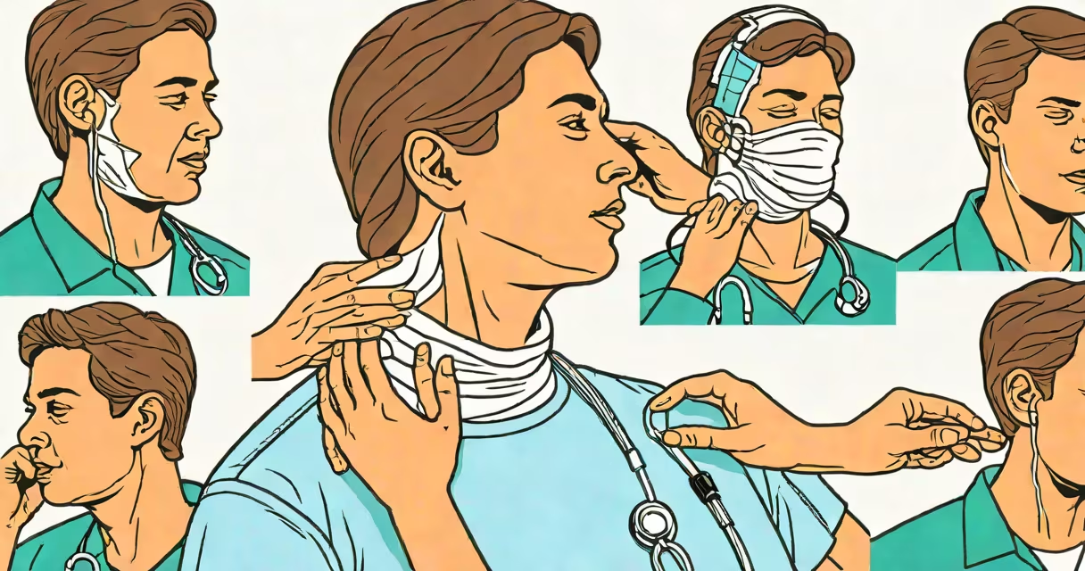
A broken neck, medically known as a cervical spine fracture, occurs when one or more vertebrae in the neck region sustain a break due to trauma. This injury requires immediate medical evaluation because it can lead to spinal cord damage, paralysis, or death if not treated promptly.
Such fractures typically result from high-impact events like car crashes, falls from height, sports injuries, or physical assaults. The cervical spine consists of seven vertebrae labeled C1 through C7, with C1 and C2 being particularly vulnerable due to their role in supporting the head and enabling movement.
According to the American College of Surgeons' Committee on Trauma, cervical spine injuries account for approximately 10% of all spinal trauma cases, with about 15% of those resulting in complete paralysis. Early recognition of signs is important to reduce morbidity and mortality.
The neck's structure allows for significant mobility but also increases risk during sudden deceleration or direct impact. When force exceeds the bone's tolerance, fractures occur. These may involve compression, burst, or flexion-distraction patterns depending on the mechanism of injury.
Spinal cord involvement is the most serious concern. Even if the bones heal, damage to neural tissue can cause permanent sensory or motor deficits. For example, a fracture at C5 may result in loss of arm function, while C6-C7 injuries often affect hand coordination and grip strength.
Imaging such as CT scans and MRI is required to confirm diagnosis. A single axial CT slice can reveal subtle fractures missed on standard X-rays, especially in unstable or complex injuries.
Signs of a broken neck do not always include visible deformity. Common indicators include:
One study published in Spine Journal found that 68% of patients with cervical spine fractures reported neck pain as the first symptom, while 42% experienced neurological deficits upon arrival at the emergency department.
Some individuals may appear stable initially but develop worsening symptoms within hours. Delayed presentation increases the risk of secondary injury due to improper movement or positioning.
Many people mistake a broken neck for a simple muscle strain or whiplash. While both can cause neck pain, a broken neck involves structural failure of bone, not soft tissue. Whiplash typically resolves within days to weeks; a fracture requires surgical intervention or immobilization.
Another misconception is that only obvious trauma leads to fracture. However, osteoporosis can weaken bones, making even minor falls in older adults sufficient to cause a break. The National Institute of Neurological Disorders and Stroke reports that vertebral fractures in adults over 65 are often underdiagnosed due to low suspicion.
Some assume that absence of pain rules out injury. But spinal cord injury can occur without acute pain-especially in patients with pre-existing conditions or those taking opioids.
Fractures are classified based on location and pattern. The most common types include:
| Type | Description | Typical Cause | Risk Level |
|---|---|---|---|
| C1 fracture (Jefferson fracture) | Burst fracture of the atlas vertebra | High-impact axial loading (e.g., diving into shallow water) | High - risk of spinal cord compression |
| C2 fracture (hangman's fracture) | Vertical split in the pedicles of C2 | Hyperextension during a fall or crash | High - instability and risk of neurologic injury |
| Compression fracture (C3-C7) | Vertebra collapses anteriorly | Flexion forces, such as rear-end car collisions | Moderate - often treatable with bracing |
Each type demands different management. For example, Jefferson fractures require strict immobilization and imaging follow-up, while compression fractures may be managed conservatively with a hard collar and physical therapy.
In 2021, a 34-year-old man was brought to the ER after a motorcycle collision. He complained of neck pain and had decreased sensation in his left arm. Initial X-ray showed no obvious fracture, but a CT scan revealed a bilateral C2 pedicle fracture consistent with hangman's fracture.
He was placed in a rigid cervical collar and underwent surgical stabilization. Without early detection, he would have faced a high risk of spinal cord injury during transport or movement.
This case underscores why clinical judgment combined with appropriate imaging is essential. Relying solely on plain films misses up to 20% of cervical fractures, according to guidelines from the Eastern Association for the Surgery of Trauma.
If someone has experienced a fall, accident, or blow to the head or neck and shows any of the following, they must go to the emergency room:
Do not attempt to move the person unless absolutely necessary. Stabilize the head and neck in neutral alignment until help arrives. Moving a person with an unstable fracture can cause irreversible spinal injury.
I once observed a paramedic team stabilize a patient with suspected cervical fracture after a skiing fall. They used a vacuum mattress and inline stabilization techniques. The patient later confirmed the injury via MRI-had movement occurred before arrival, the outcome could have been devastating.
One overlooked fact is that signs of a broken neck may not appear immediately. Delayed onset of symptoms-such as progressive weakness or difficulty walking-can occur within 24 hours post-injury. This delay is due to swelling or shifting fragments compressing nerves over time.
Also, patients with altered mental status (from intoxication or concussion) may not report pain accurately. In these cases, physical exam findings like asymmetrical reflexes or abnormal gait become critical diagnostic clues.
For example, a patient who cannot touch their chin to their chest despite neck pain may have a fracture. The inability to perform this simple motion-a test called the "chin-to-chest" maneuver-is a red flag in trauma assessment.
If you suspect a broken neck, call emergency services immediately. Do not attempt to move the person. Keep them still, support the head and neck in alignment, and wait for trained professionals.
Once stabilized, imaging protocols follow established guidelines. The Canadian C-spine Rule and NEXUS criteria help determine which patients need imaging based on risk factors like age, mechanism of injury, and neurological status.
Early diagnosis reduces complications. A 2020 review in Journal of Trauma and Acute Care Surgery found that patients evaluated within 1 hour of injury had a 30% lower rate of long-term disability compared to those seen after 4 hours.