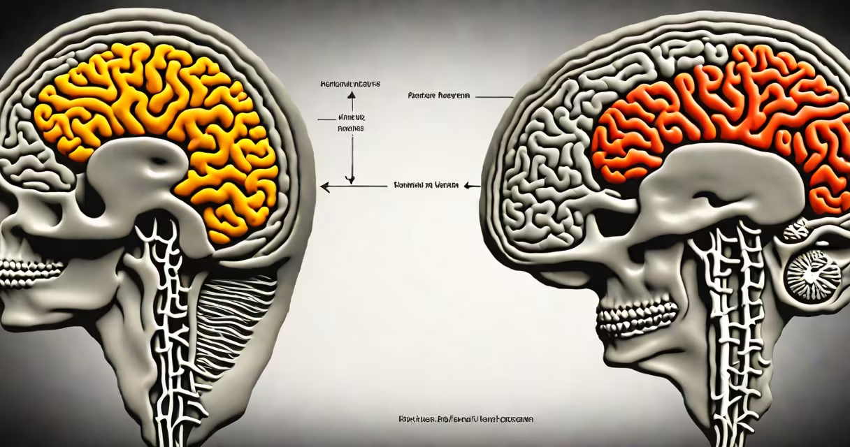
Depression alters brain structure and function through measurable changes in size, connectivity, and chemistry. Key regions like the hippocampus shrink, neural circuits involved in mood and decision-making show disrupted activity, and neurotransmitter systems such as serotonin and dopamine become imbalanced. These shifts contribute to persistent low mood, impaired concentration, and reduced motivation.
Neuroimaging studies confirm that depression isn't just emotional-it's biological. Structural MRI scans reveal consistent reductions in gray matter volume in areas responsible for emotion regulation and memory. Functional MRI shows abnormal patterns of activation during emotional processing, especially in the prefrontal cortex and limbic system. These findings are not hypothetical-they're documented in peer-reviewed research from institutions like the National Institute of Mental Health and Harvard Medical School.
The brain changes seen in depression are not simply side effects. They represent core features of the illness. When left untreated, these alterations may persist even after mood symptoms improve, increasing the risk of relapse. Early intervention can halt or reverse some of this damage, underscoring the urgency of timely treatment.
Brain imaging reveals that chronic depression leads to measurable shrinkage in specific regions. The hippocampus-a structure critical for memory formation and stress regulation-loses volume over time. A 2016 study published in *JAMA Psychiatry* found that individuals with recurrent major depressive disorder had hippocampal volumes 4% smaller on average than matched controls. This reduction correlates with longer illness duration and poorer response to antidepressants.
Another region affected is the anterior cingulate cortex (ACC), which modulates attention and emotional responses. Reduced activity here is linked to difficulty shifting focus away from negative thoughts-a sign of rumination in depression. fMRI scans show that people with depression exhibit hyperactivity in the amygdala, the brain's fear center, while underactivating the dorsolateral prefrontal cortex, which normally helps regulate emotional reactions.
These shifts aren't reversible overnight. A longitudinal study tracking patients over five years showed that those who experienced multiple depressive episodes had progressive gray matter loss in the prefrontal cortex. The brain adapts to prolonged stress by reducing synaptic density and altering neuroplasticity-the ability to form new connections. This creates a feedback loop: poor mood impairs brain function, which worsens mood.
Depression involves dysregulation of key neurotransmitters: serotonin, norepinephrine, and dopamine. Serotonin influences mood, sleep, and appetite. Low levels are associated with persistent sadness and irritability. Norepinephrine affects alertness and energy; its depletion contributes to fatigue and mental fog. Dopamine, tied to reward and motivation, drops in anhedonia-the inability to feel pleasure.
But the picture is more complex than "low serotonin." Research from the University of California, San Diego, demonstrates that serotonin receptors in depressed individuals often become less sensitive, meaning even normal levels fail to trigger proper signaling. This receptor desensitization explains why some patients don't respond to SSRIs (selective serotonin reuptake inhibitors), which work by increasing available serotonin but cannot restore receptor function.
Dopamine pathways also suffer. PET scans show reduced dopamine transporter availability in the striatum-the brain's reward hub-among people with severe depression. This deficit aligns with clinical observations: patients report losing interest in hobbies, socializing, or even basic self-care. The brain's reward system becomes effectively "shut down" by prolonged stress and inflammation.
Depression doesn't affect the brain uniformly. Different subtypes correlate with distinct neural patterns. For example:
| Type | Key Brain Changes | Associated Symptoms |
|---|---|---|
| Atypical Depression | Hyperactive hypothalamus, increased cortisol release | Weight gain, excessive sleep, carbohydrate cravings |
| Psychotic Depression | Reduced connectivity between prefrontal cortex and thalamus | Hallucinations, delusions, disorganized thinking |
| Treatment-Resistant Depression | Severe hippocampal atrophy, low BDNF levels | Persistent anhedonia, suicidal ideation despite medication |
These differences explain why treatments vary. Atypical depression responds better to medications that boost both serotonin and norepinephrine. Psychotic depression often requires antipsychotics alongside antidepressants. Treatment-resistant cases may benefit from ketamine or transcranial magnetic stimulation (TMS), which directly stimulate underactive brain regions.
Many assume depression is purely psychological or a sign of weakness. That view ignores decades of neuroscientific evidence. Brain changes in depression are not imagined-they're visible on scans and reproducible across populations. A 2020 meta-analysis in *The Lancet Psychiatry* confirmed that structural brain differences in depression are statistically significant and clinically relevant.
Another misconception is that recovery means the brain returns to normal. While many patients regain function after treatment, some structural changes persist. A 2018 study found that 30% of individuals who recovered from major depression still showed reduced hippocampal volume compared to never-depressed controls. This suggests lasting neurological impact-even when symptoms disappear.
Some also believe antidepressants fix the brain. While they help restore balance, they don't reverse all physical changes. Antidepressants primarily modulate neurotransmitter levels, not brain structure. Long-term recovery often requires behavioral therapy, lifestyle changes, and sometimes neuromodulation techniques.
A 2021 study tracked 120 adults diagnosed with moderate to severe depression using serial MRI scans and symptom assessments. Participants received either escitalopram (an SSRI) or placebo over 12 weeks. Those on medication showed a 3.2% increase in hippocampal volume by week 12, while the placebo group lost 1.8%. Simultaneously, their Beck Depression Inventory scores dropped by 54%, compared to 22% in the placebo group.
The brain changes preceded symptom improvement. Hippocampal growth began at week 4, before mood scores shifted. This suggests that neurobiological recovery may precede subjective feeling better-an insight important for clinicians managing treatment expectations.
If you're struggling with persistent low mood, fatigue, or loss of interest, seek evaluation from a mental health professional. Early diagnosis and treatment reduce long-term brain impact. Options include medication, psychotherapy, lifestyle interventions, and brain stimulation therapies.
Understanding what depression does to the brain removes stigma and clarifies treatment goals. It's not a flaw in character-it's a medical condition with tangible effects on brain biology. Recognizing this enables better decisions, faster recovery, and reduced risk of recurrence.