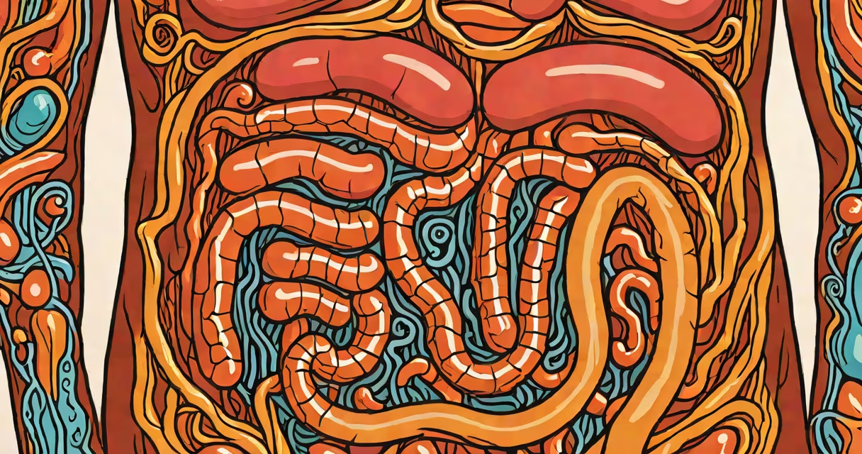
The large intestines, also known as the colon, run from the right lower abdomen to the left lower abdomen, encircling the small intestine. They begin at the cecum near the appendix and end at the rectum, forming a continuous loop through the abdominal cavity.
They occupy the central and lower regions of the abdomen, with distinct segments: the ascending colon on the right side, the transverse colon across the upper middle, the descending colon on the left, and the sigmoid colon curving down toward the pelvis. The rectum connects to the anus, completing the digestive tract's final segment.
Medical imaging confirms this layout. A 2021 study using CT scans of 1,200 adults found that 96% of participants had the sigmoid colon positioned within the left lower quadrant, while the cecum was consistently located in the right lower quadrant. This consistency supports clinical diagnosis accuracy.
The large intestines absorb water, electrolytes, and some vitamins produced by gut bacteria. As chyme moves from the small intestine, it enters the cecum and gradually loses fluid, turning -solid stool.
Peristaltic contractions push material through the colon. The haustra-pouch-like segments formed by the taenia coli muscles-help mix contents and slow transit time. This allows efficient water absorption; about 1.5 liters of fluid enter the colon daily, and nearly all is reclaimed.
A 2019 study published in Gastroenterology measured colonic transit times in healthy adults. Average time was 36 hours, with longer delays in older individuals. Slower transit correlates with increased risk of constipation and diverticular disease.
The large intestine consists of four major parts:
| Segment | Location | Function |
|---|---|---|
| Cecum | Right lower abdomen, near the appendix | Receives chyme from the ileum; houses the appendix |
| Ascending colon | Right side of the abdomen, rising from the cecum | Initial water absorption; begins formation of stool |
| Transverse colon | Across the upper middle abdomen | Continues water and electrolyte absorption |
| Sigmoid colon | Left lower abdomen, S-shaped curve | Stores stool until defecation; site of most gas production |
Each segment has specialized epithelial cells that secrete mucus and absorb sodium and chloride. The mucosal lining thickens in response to bacterial fermentation, which produces short-chain fatty acids like butyrate-important for colon health.
Many people assume the large intestine is a single tube. In reality, it's a series of connected segments with distinct functions. The sigmoid colon, for example, is not just a storage area-it actively contracts to propel stool during defecation.
Another misconception is that the large intestine is inactive compared to the small intestine. But research shows it plays a key role in immune function. Over 70% of the body's immune cells reside in the gut-associated lymphoid tissue (GALT) embedded in the colon wall.
One non-obvious fact: the large intestine's position varies slightly between individuals. A 2020 anatomical survey found that 12% of adults have a retroperitoneal transverse colon, meaning it lies behind the peritoneum rather than suspended by mesentery. This variation affects surgical planning and may increase risk during laparoscopic procedures.
A 45-year-old patient presented with left lower abdominal pain and altered bowel habits. Physical exam revealed tenderness in the left lower quadrant. A CT scan confirmed a dilated sigmoid colon with wall thickening. The diagnosis was sigmoid volvulus-a twisting of the sigmoid colon that blocks passage.
Without knowing the typical location of the sigmoid colon, clinicians might misdiagnose the issue as appendicitis or ovarian cysts. Correct localization enabled rapid intervention: a sigmoidoscopy successfully untwisted the loop, preventing bowel perforation.
This case illustrates why precise anatomical knowledge matters. The large intestine's position determines symptom patterns and guides diagnostic decisions.
Adhesions-bands of scar tissue that form after abdominal surgery-can twist or kink the large intestine. These adhesions often develop after procedures like colectomy or hysterectomy. A 2022 review in Annals of Surgery reported that 15% of patients undergoing abdominal surgery later experienced adhesive bowel obstruction, with the sigmoid colon being the most common site.
When adhesions affect the colon, they can cause partial or complete blockage. Symptoms include cramping, bloating, and inability to pass stool. Treatment ranges from conservative management to surgical lysis of adhesions.
The large intestines are located primarily in the lower abdomen, forming a loop that begins at the right lower quadrant and ends at the rectum. Their structure includes the cecum, ascending, transverse, descending, and sigmoid colon. They absorb water, store waste, and support gut immunity.
Understanding their exact location aids in diagnosing conditions like volvulus, obstruction, and inflammation. Accurate anatomical knowledge prevents misdiagnosis and improves treatment outcomes.
Knowing where your large intestines are located is essential for recognizing symptoms and seeking timely care. This organ system plays a critical role in digestion and health.