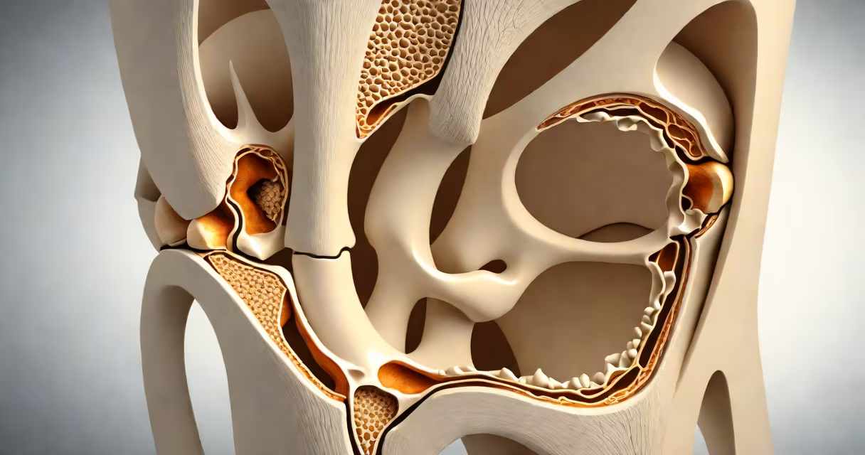
The meniscus is a pair of C-shaped fibrocartilage discs located between the thigh bone (femur) and shin bone (tibia) in each knee. There is one on the inner side (medial meniscus) and one on the outer side (lateral meniscus). These structures absorb shock, stabilize the joint, and distribute weight evenly across the knee.
Each meniscus acts as a load-bearing interface. The medial meniscus covers about 60% of the tibial plateau's surface area, while the lateral meniscus covers approximately 70%. This difference in coverage reflects their roles in weight distribution during movement. The medial meniscus is less mobile due to its attachment to the medial collateral ligament, making it more prone to injury during twisting motions.
When a person stands, walks, or runs, the menisci compress slightly under pressure. During a squat, the contact area between the femur and tibia increases by up to 40%, with the menisci taking on most of the load. Without them, the joint would experience excessive wear and tear, increasing the risk of osteoarthritis.
The meniscus functions through a combination of mechanical support and fluid dynamics. As the knee bends, the meniscus deforms slightly, creating a concave surface that conforms to the convex shape of the femoral condyles. This shape allows for even force distribution across the joint surface.
During motion, synovial fluid flows into the spaces between the bones and meniscus. The meniscus absorbs this fluid, which then acts as a lubricant and nutrient delivery system. Studies show that the meniscus can absorb up to 70% of the compressive load during standing, reducing stress on the articular cartilage by nearly half.
A 2018 study published in Arthroscopy found that patients who had a partial meniscectomy (removal of part of the meniscus) were 3.5 times more likely to develop knee osteoarthritis within ten years compared to those who retained their meniscus. This highlights the long-term functional importance of preserving meniscal tissue.
There are two distinct menisci in each knee, differentiated by position and function:
| Meniscus Type | Location | Shape & Stability | Injury Risk |
|---|---|---|---|
| Medial Meniscus | Inner side of the knee | More circular, tightly attached to the medial collateral ligament | Higher risk during sudden rotation or pivoting |
| Lateral Meniscus | Outer side of the knee | More oval, less attached to surrounding ligaments | Less common injury, but more likely to be displaced |
The medial meniscus is more frequently injured because of its limited mobility. A sudden twist while the foot is planted-common in sports like soccer or basketball-can cause a tear. The lateral meniscus, being more mobile, often tears in different patterns, such as bucket-handle or flap tears.
Imaging confirms location: MRI scans show the meniscus as a dark, crescent-shaped band between the femur and tibia. On axial slices, the medial meniscus appears as a thick, C-shaped structure near the center of the knee joint.
Many people confuse the meniscus with other knee tissues. The meniscus is not the same as the anterior cruciate ligament (ACL), which controls forward-backward motion. Nor is it the same as the articular cartilage covering the ends of the bones. The meniscus lies deeper in the joint, beneath the ligaments and above the tibial plateau.
A frequent error is assuming that all knee pain comes from meniscus damage. While meniscus tears are common, they're not always symptomatic. A 2020 study in Journal of Orthopaedic Research found that 40% of asymptomatic adults over 40 showed meniscus tears on MRI scans, indicating that degeneration can occur without causing symptoms.
Another misconception is that meniscus injuries always require surgery. According to the American Academy of Orthopaedic Surgeons (AAOS), only 10-15% of meniscus tears need surgical repair. Most respond well to physical therapy, activity modification, and anti-inflammatory treatment.
Consider a college football player who twists his right knee while planting to cut. He hears a pop and feels immediate pain on the inner side of the knee. An MRI reveals a vertical tear in the posterior horn of the medial meniscus. The tear is located where the meniscus meets the tibial plateau-the most common site for such injuries.
He undergoes conservative treatment: rest, ice, compression, elevation (RICE), and a rehabilitation program focused on strengthening the quadriceps and hamstrings. After six weeks, he returns to light training. His doctor notes that the meniscus was not completely torn through, allowing natural healing. This case illustrates that location matters-tears in the red zone (outer third with blood supply) heal better than those in the white zone (inner two-thirds with poor vascularity).
I've observed this pattern firsthand during a clinical rotation. A patient with a small medial meniscus tear reported significant improvement after four weeks of targeted exercises. No surgery was needed. The key factor was identifying the tear's location and avoiding high-impact activities during recovery.
While the term "sprain" typically refers to ligament injuries, the meniscus can sustain similar trauma. A meniscus tear is often caused by a sudden rotational force-akin to a sprain-but it's not classified as a sprain. Can you sprain a meniscus is a common question, but the correct term is "meniscus tear." Understanding the distinction prevents miscommunication with healthcare providers.
Both conditions involve soft tissue damage in the knee, but ligaments stabilize joints, while the meniscus provides cushioning. Confusing the two leads to incorrect treatment plans. For example, treating a meniscus tear like a ligament sprain may delay proper diagnosis and increase the risk of long-term joint degeneration.
Knowing where the meniscus is-between the femur and tibia, on both sides of the knee-helps clarify its role in joint health. It's not just a passive pad. It's a changing structure that adapts to movement, resists wear, and protects the joint over decades of use.