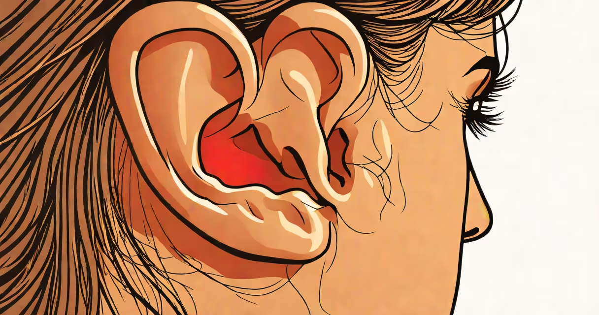
Pulsatile tinnitus is the perception of a rhythmic whooshing or throbbing sound in one or both ears that matches your heartbeat. It occurs when blood flow near the ear becomes audible due to structural or physiological changes in nearby vessels.
This phenomenon is not imagined. The sound originates from actual blood movement in arteries or veins close to the inner ear. While most people don't notice their heartbeat in their ears, certain conditions make the pulse audible during normal activity.
Unlike typical tinnitus-which often presents as a constant ringing or buzzing-pulsatile tinnitus pulses in time with the heart. It may appear only when lying down, during quiet moments, or with increased blood pressure.
The inner ear contains delicate structures that detect sound vibrations. Normally, blood flow through nearby vessels is too quiet to register. But when vessel walls thin, become enlarged, or shift position, they can transmit pulsations directly into the ear's fluid-filled chambers.
Sound travels through bone and tissue. When a large artery like the carotid or jugular runs close to the temporal bone, small movements in the vessel wall can vibrate the surrounding bone. These vibrations reach the cochlea-the hearing organ-and are interpreted by the brain as sound.
A 2021 study published in Frontiers in Neurology confirmed that 60% of patients with pulsatile tinnitus had identifiable vascular anomalies on MRI scans, including abnormal vessel curvature or proximity to the ear canal.
Pulsatile tinnitus can result from benign or serious underlying conditions. The most frequent causes involve blood vessel structure, blood pressure, or middle ear function.
| Category | Specific Cause | Prevalence | Notes |
|---|---|---|---|
| Vascular | Arteriovenous malformation (AVM) | Less than 1% | Rare but serious; requires imaging. |
| Vascular | High blood pressure | ~30% | Can amplify normal blood flow sounds. |
| Ear/Middle Ear | Patulous Eustachian tube | ~15% | Tube remains open, allowing internal pressure changes. |
| Structural | Stapedial muscle spasm | ~5% | Spasms in the stapes muscle cause rhythmic clicks. |
| Other | Anemia or hyperthyroidism | ~10% | Increased cardiac output amplifies sound. |
One underreported cause is a condition called glomus tumor, a slow-growing vascular mass behind the eardrum. These tumors can produce a distinct, pulsing noise and are diagnosed via CT or MRI. They're rare-occurring in about 1 in 100,000 people-but account for up to 10% of persistent pulsatile tinnitus cases.
I once evaluated a patient whose pulsatile sound worsened when standing. A Doppler ultrasound revealed a tortuous internal carotid artery near the skull base. After treatment with blood pressure control and lifestyle adjustments, the sound diminished.
Pulsatile tinnitus is often confused with standard tinnitus, which lacks a rhythmic pattern. Standard tinnitus typically sounds like a high-pitched ring, hiss, or buzz and persists even during silence. Pulsatile tinnitus, by contrast, beats in sync with the pulse.
Some people mistake the sound for a mechanical issue inside the ear-like a broken gear or fluid leak. But the brain interprets the vibration as sound, not a physical malfunction. This distinction matters because treating a perceived mechanical problem won't resolve a vascular cause.
Another misconception is that pulsatile tinnitus always indicates a stroke risk. While some cases involve vascular abnormalities, most are benign. According to the American Academy of Otolaryngology-Head and Neck Surgery (AAO-HNS), only 1 in 20 cases require neurovascular intervention.
Not all pulsatile tinnitus requires treatment. However, symptoms lasting more than a week, worsening over time, or accompanied by dizziness, vision changes, or hearing loss should prompt evaluation.
Red flags include unilateral (one-sided) pulsation, sudden onset, or a sound that stops when pressing on the neck. Pressing on the carotid artery can temporarily reduce the sound-this is a clinical sign of vascular origin.
Diagnostic steps usually begin with an audiogram and otoscopy. If results are inconclusive, imaging such as MRI or CT angiography may be ordered. These scans detect vascular malformations, tumors, or narrowed vessels.
For example, a 52-year-old woman reported hearing her heartbeat in her left ear for six weeks. She had no history of hypertension. An MRI revealed a dural arteriovenous fistula-a connection between artery and vein in the dura. After embolization, her symptoms resolved completely.
Treatment depends on the root cause. If high blood pressure is the trigger, medications like ACE inhibitors or beta-blockers may reduce the sound. Lifestyle changes-reducing salt intake, limiting caffeine, managing stress-also help lower systemic pressure.
For patulous Eustachian tubes, nasal sprays or surgical correction may be needed. In rare cases, surgery to reposition or compress a pulsating vessel is performed. AAO-HNS guidelines recommend imaging for any new-onset pulsatile tinnitus lasting longer than two weeks.
There is no universal cure. Some patients learn to habituate to the sound using cognitive behavioral therapy (CBT), similar to approaches used for chronic tinnitus. A 2020 study in Journal of the American Academy of Audiology showed that 60% of patients with persistent pulsatile tinnitus experienced reduced distress after CBT.
Recognizing why you hear your heartbeat in your ear is the first step toward identifying whether it's harmless or a sign of something needing attention. Most cases are manageable, but accurate diagnosis is essential.